Optimising photosynthesis by altering the structure of leaves
Project facts:
- Start date: 1st April 2012
- End date: 1st April 2015
- Funder: BBSRC
- Joint award with University Of Sheffield
- Investigators:
Prof Andy Fleming (PI)
Prof Sacha Mooney
- Researcher:
Dr Radoslaw Pajor
Background
With growing human population and losses in the air and soil quality, the food security becomes a major concern and challenge. The soil structure and its effects on soil properties and ecosystems are widely explored. However, with crops such as wheat, rice and maize being the providers of over 50% of worlds diet, the lacking information is whether the plants themselves are ready to withstand environmental difficulties such as increasing CO2 concentration.
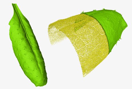
3D visualization of whole leaf (left) and segmented intracellular pore space from the tip of the Arabidopsis leaf (right). Pore space (yellow) was illustrated by peeling the plant tissue (green) away.
Survival of the plants and securing the yields rely on optimal process of photosynthesis. One of the main factors regulating photosynthetic performance is the availability of CO2. The CO2is used in processes occurring inchloroplasts, inside the cells. However, the gas enters the leaf inter-cellular pore-space through stomata and has to diffuse through the leaf to the target cells. The process of diffusion is limited by stomatal conductance (gm) which depends mainly on the arrangement of mesophyll. However, the exact link between the structure of plant tissues and photosynthetic efficiency remained unclear. Project
The gaps in the knowledge were mainly due to the lack of suitable methods for non-invasive visualisation and quantification of plant tissues in 3D. Most of the methods used in classical histology were time-consuming, destructive and most importantly limited to 2D so unable to capture the 3D complexity of leaf architecture and link it to plant physiology.
The state of art high-resolution, X-ray tomography combined with image analysis methods allows visualising and quantifying the morphology of the whole leaf at the resolution (voxel size) of 15 µm, as well as exploring the structural descriptors of plant tissues at 2.75 µm. Structural analysis provides detailed information about the pore networks such as pore volume, pore size distribution, connectivity, surface area and shape descriptors in 3D. X-ray CT is non-invasive technique, therefore structural measurements are performed on the very same aerial plant organs which were subject to analysis capturing physiological performance of the plants.
Screening many genotypes and combining the structural descriptors with measurements of plant physiology leads to a better understanding of the relations between plant tissue structure and functional requirements for design of future-proof leaves.