Micro Computed Tomography (MicroCT) for Food Technology
What is CT?
CT is a technology which allows you to see inside an object in 3D without cutting it open. Materials with different densities can be separated, false coloured and numerically measured. Through using the latest equipment, we are able to acquire images at resolutions from 150 µm down to <1µm.
Examples of CT in Food Technology
A journey through a chocolate digestive biscuit
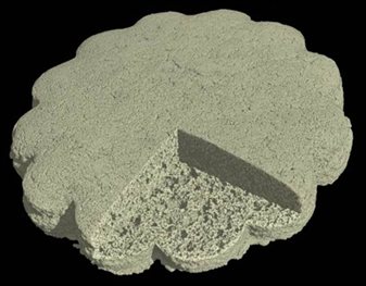
Discovering the secrets of bread crispiness
Revealing the complexity of multi-layered chocolate
The following schematic illustrates how a food product such as this aerated chocolate could be numerically quantified:
How do food additives affect product texture?
Just by changing the flavour solvent in this shortcake biscuits from propylene glycol to triacetin, the microstucture changes drastically. The biscuits with p ropylene glycol had smaller pores and higher porosity than those made with triacetin which may be influence the generation and stability of biscuit aroma compounds (see article for more details).
How can the technology help you?
The technique has a number of advantages over current methods of analysis such as optical and electron microscopy:
- Food products can be investigated in their natural state at atmospheric pressure and temperature. Other methods such as electron microscopy require a vacuum and coating of the sample.
- Microscopy requires the product to be mechanically sectioned into thin slices. Even the most careful preparation can have destructive effects on the product. In contrast, X-ray tomography can visualise the internal structure in 3D without any cross-sectioning or preparation. Reliable 2D and 3D information can then be derived from the data.
- Microscopy is restricted to analysing small sections of the overall product. A large number of repeats are required in order to give statistically relevant results. With X-ray tomography, the entire product can be imaged and analysed, reducing the number of repeats that are needed.
- X-ray tomography data can be acquired at relatively high spatial resolutions ranging from <1µm to 150µm depending on the product’s size.
Food quality can be explored visually and quantitatively in 3D, studying characteristics such as:
- Porosity e.g. to investigate food texture.
- Distribution and size of particles such as salt crystals or bubbles.
- Macro and microstructure (for structural and mechanical properties e.g. deformation, fracture, homogeneity).
- The 3D location and morphology of various ingredients which can be distinguished from one another when they have significantly different densities.
- Thickness of material layers and other geometrical measurements can be made.
Can we help you?
Using our interdisciplinary team of experts we are able to take care of the whole CT data acquisition and data analysis process for you. We will take time to understand your research questions and discuss how we might be able to answer them. Through designing a customised data acquisition and image analysis package for your problem, you will obtain the best solution that X-ray micro-tomography can offer.
For more information or to discuss a possible project contact Craig Sturrock: