Cardiology Teaching Package
A Beginners Guide to Normal Heart Function, Sinus Rhythm & Common Cardiac Arrhythmias
Sinus Rhythm
Sinus rhythm is the name given to the normal rhythm of the heart where electrical stimuli are initiated in the SA node, and are then conducted through the AV node and bundle of His, bundle branches and Purkinje fibres.
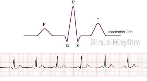
Depolarisation and repolarisation of the atria and ventricles show up as 3 distinct waves on ECG. A unique labelling system is used to identify each wave.
Although the diagram shows 5 waves, we will concentrate on 3 waves. You will not always see a Q wave or an S wave on an ECG.
This is why only 3 waves are emphasised when you are learning from scratch.
Image: P,QRS & T Wave
Remember what was said earlier about the electrical stimuli passing through myocardial cells. The more cells there are, the more voltage is required. Now look at the previous pictures of the heart. The walls of the atria are much thinner and smaller than those of the ventricles.
Less muscle means less cells which means less voltage.