Non-invasive assessment of portal hypertension using quantitative magnetic resonance imaging
In patients with cirrhosis, the development and progression of portal hypertension is related to worse outcomes. However, the standard technique of assessing portal pressure is invasive and not widely used in clinical practice. Here, we have studied the use of non-invasive MRI in evaluating portal pressure. The MRI measures of liver architecture and blood flow in the splenic artery correlated well with portal pressure. Therefore, this non-invasive method can potentially be used to assess portal pressure in clinical trials and monitoring treatment in practice.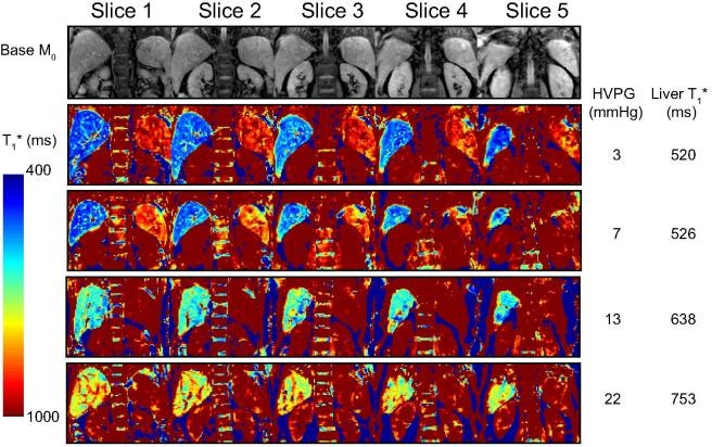
Figure: Example of T1 relaxation maps. Top row shows example image quality of the coronal-oblique imaging slices for base equilibrium M0 data acquired from a patient with HVPG of 3 mmHg. Subsequent rows show example coronal-oblique bFFE T1∗ maps showing the liver and spleen, together with HVPG (mmHg) and mode of the liver T1∗ value (ms) across a range of HVPG from 3–22 mmHg, with columns showing each of the five slices collected for the T1∗ map.