Advanced imaging analysis and Machine Learning
Automated Segmentation of the Kidneys
Segmentation of kidneys is an important yet time consuming aspect of many studies. Existing automated segmentation methods using classical image processing are specific to a single pathology. Using a convolutional neural network the kidneys of both a healthy control and chronic kidney disease cohort can be segmented with better than human precision. This work has been published in
Magnetic Resonance In Medicine and the trained convolutional neural network is available as an easy to use
GUI,
Python package or part of the
UKRIN Kidney Analysis Toolbox (UKAT).
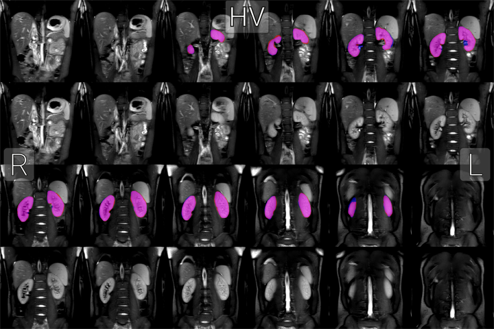
The kidneys of a healthy volunteer segmented by a human (blue overlays) and automatically (red overlays) and the overlap between the two (magenta overlays).
In addition to being used on data from Philips scanners here in Nottingham, the accuracy this technique has been extensively verified on multi-vendor data and as such is being used as part of both the AFiRM and CMORE projects. These multi-site, multi-vendor studies involve the analysis of data from hundreds of volunteers, as such the automation of renal segmentation is invaluable.
Work is now focusing on automated sementation of the renal cortex and medulla.
Layer Based Analysis of the Kidneys
Most analysis of renal MRI data is based on defining regions of interest (ROI) for different tissues in the kidneys however this technique can be variable between investigators. To eliminate the subjectivity in ROI definition, the distance from each voxel to the surface of the kidney is calculated allowing the kidney to be divided into layers and quantitative calculated allowing the kidney to be divided into layers and quantitative maps to be expressed as a function of renal depth.