A Nephrectomy model to understand renal microstructure
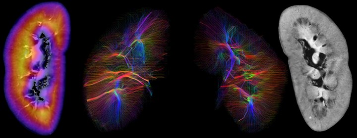
The study of post-mortem brain tissue using MRI has been shown to provide a useful tool to assess whole organ microstructure and pathology with high spatial resolution. A clear understanding of the effects of fixation on the tissues MRI parameters is crucial for interpreting ex-vivo MRI studies however this understanding did not exist for renal tissue. A protocol for consistent scanning of formalin fixed ex-vivo kidneys has been developed and was presented at ISMRM 2019.