Cardiology Teaching Package
A Beginners Guide to Normal Heart Function, Sinus Rhythm & Common Cardiac Arrhythmias
Atrial Tachycardia
Image: Sinus Tachycardia (close up)
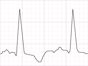
Although the rate is 140 bpm, it is possible to clearly identify the P wave, QRS complex and T wave.
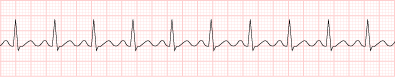
Image: Sinus Tachycardia (full strip)
Atrial Tachycardia
Image: Atrial Tachycardia (multi-focal)
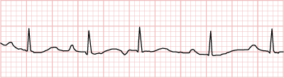
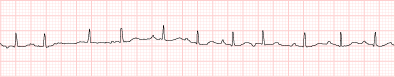
Image: Atrial Tachycardia (full strip)
Note in this rhythm how the P wave is constantly changing shape. If the P wave is not a nice, rounded shape, often this will indicate an atrial rhythm, where the stimulus for depolarisation is coming from a focus other than the SA node.
In the example above there are at least 3 different areas (foci) stimulating the atria, as there are at least 3 different shaped P waves.