Cardiology Teaching Package
A Beginners Guide to Normal Heart Function, Sinus Rhythm & Common Cardiac Arrhythmias
The U Wave
Finally, it is possible to see another wave after the PQRST complex. This is known as a U wave. It is not very common and is easy to overlook. (see example below.)
Image: Image: U waves are indicated by the arrows
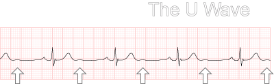
In a normal heart beat, the T wave represents repolarisation of the ventricles, specifically the repolarisation of the AV node and bundle branches. The U wave occurs when the ECG machine picks up repolarisation of the Purkinje fibres.
A "U wave" can also occur with electrolyte imbalances (potassium) but, again, this is not very common.
Authors Note:
I had never seen a naturally occurring U wave in 6 years in Cardiology, or maybe just never noticed. But, as fate would have it, when preparing this section I came across 3 patients with U waves on the same day.