The HS-PrediCt Blog
Brainbox visit and TMS equipment arrival
20th October 2022
Alexina Whitley, Eszter Porter and Muzna Shehzad
In June the HS-PrediCt team had an exciting delivery from our friends at Brainbox (
https://brainbox-neuro.com/) – the arrival of a Transcranial Magnetic Stimulation (TMS) and Neuronavigation System. Our Principal Investigator, Dr Lauren Hadley, won the use of this equipment for a few months after winning the Brainbox Initiative (
https://brainbox-initiative.com/) Research Challenge in 2019. Dan Phillips, Co-Founder and Director of Brainbox Ltd, delivered the equipment to us and kindly stayed to help set-up in the lab and train the team on how to use the system.
What is TMS and Neuronavigation?
Transcranial Magnetic Stimulation (TMS)
TMS is a non-invasive brain stimulation method commonly used in research to help us better understand the role of different brain areas in various tasks. TMS can be used to disrupt neural activity and measure the consequences that this has on task performance. It can also be used to increase activity in specific regions, for example raising motor activity above threshold to make it easier to measure activation from muscles. A TMS system generally consists of the stimulator, and a coil that is placed on the participants’ scalp so that its centre is over the brain area you are aiming to stimulate. Different types of stimulators and coils exist for different purposes, but in this blog, we’ll stick to what we’ll be using in our lab.
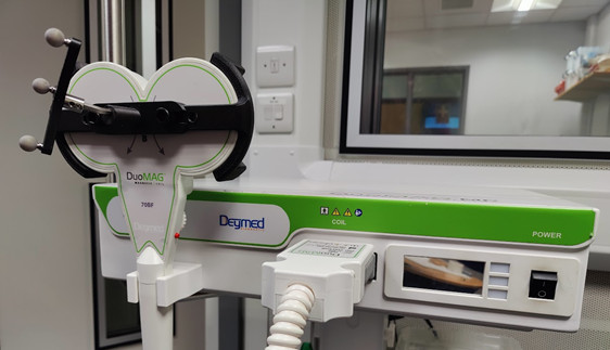
Our TMS System
So, how does TMS work?
In simple terms, the TMS coil contains windings of copper. The stimulator that is used to deliver a “pulse” via the TMS coil passes a brief current through these copper windings, which in turns leads to a brief magnetic field being generated from the centre of the coil. This varying magnetic field stimulates nerve cells in the region of the brain under the coil– hence being called transcranial magnetic stimulation. As mentioned earlier, there are different types of TMS systems. The one used in our lab is single pulse TMS, in which one pulse is delivered every few seconds.
Is TMS safe?
When first hearing about TMS, it may sound somewhat scary – but it’s a safe tool to use (of course when following the recommended safety guidelines (Rossi et al., 2009; 2021). The first TMS device was developed in 1985 by Anthony Barker and colleagues in Sheffield, and after being around for decades there has been no significant long-term side effects of TMS reported. Most people only feel a sort of tapping sensation on their head when a TMS pulse is delivered. TMS carries some very small risks, though in extremely rare cases seizures have been reported (less than 1 in every 30, 000 sessions; Rossi et al., 2009, Lerner et al., 2019). However, as we are following the latest safety guidelines and using the safest type of stimulation, we do not anticipate such adverse effects.
Neuro-navigation System
A neuronavigational system allows for accurate and reliable TMS administration. The system includes an infrared camera that tracks the location of TMS coil in relation to the participants’ head using reflective reference balls. Sets of three reference balls are attached to the participants head and to the TMS coil. The associated software allows researchers to mark where the coil should be held in relation to the participant’s scalp to stimulate the target area, and gives feedback throughout the experiment on coil orientation, angle, and placement – all these things are important to ensure you’re stimulating exactly where you planned to and get the best results.

Three metal spheres (trackers) attached to the TMS coil and participants head
This is a really cool (and dare we say – fun) piece of equipment to help ensure the most accurate and reliable brain stimulation. It takes a bit of practice to keep a steady hand and get to grips with the software, so the HS-PrediCt team are very grateful to all our lovely colleagues and lab mates who have donated their time and heads to our practice and training!
Our TMS Research
What will we be researching using TMS?
We are using TMS to investigate the mechanism by which we predict upcoming speech. In particular, we are interested in better understanding any potential differences in predictive mechanisms between those with hearing impairment and those with normal hearing.
Previous research suggests that the same mechanisms that are required for producing speech are also involved in the processing of speech (Wilson et al., 2004; Fadiga et al., 2002).
In other words, when listening to a conversational partner, individuals use their motor system to covertly simulate the other speaker’s utterances. This motor activation is speech specific, recruiting areas required for the specifically relevant mouth movements (i.e., motor regions for controlling lip movements are involved in perception of speech sounds that involve the lips (/ba/pa/); Möttönen & Watkins, 2009). In different instances, we may use simulation to a greater degree/ extent. For example, when the environment is noisy, we may use prediction to compensate for the increased difficulty. In fact, there is some evidence that the areas of the motor system required for producing speech are active during speech perception (Wilson et al., 2004, Bartoli et al., 2015), particularly during degraded listening situations (e.g., when listening to speech in noise versus without noise; Murakami et al., 2011). This increase in motor activity when listening to speech in noise suggests motor activation may be important for successful comprehension when listening is challenging.
However, some research suggests that there is breakdown in this mechanism of simulation for those with hearing impairment. For instance, eye-tracking experiments have shown people with hearing loss are slower at processing sentences (Wendt et al., 2015). Additionally, older adults with hearing loss show reduced motor activation when listening to speech (Panouillères & Möttönen, 2018). We aim to examine speech-related motor activity when older adults with normal hearing and hearing loss listen to speech. The purpose of this research will be to determine whether there are differences between people with normal hearing versus impaired hearing in the way that the motor system is utilised for predicting upcoming speech.
How are we using TMS?
We will be using single-pulse TMS to investigate the motor system. The motor system consists of both the central nervous system (CNS) and peripheral nervous system (PNS) and supports the body’s movement and movement planning. The primary motor cortex is handily one of the easiest places you can stimulate, due to it being part of the cerebral cortex and not a deeper structure. Target locations for stimulation that are closer to the scalp, allow for more accurate stimulation and lower stimulator intensities to be used (which also helps reduce current spread, meaning you’re less likely to also stimulate surrounding structures).
In our research, we want to investigate the level of excitability in the lip motor region during speech perception (i.e., how much the lip region of the brain is being utilised during speech listening). We will therefore be using electromyography (EMG) alongside the TMS. EMG involves using surface electrodes to measure electrical signals associated with the contraction of a muscle (in this case the lip). You can see an example of the electrodes being worn by a colleague below.
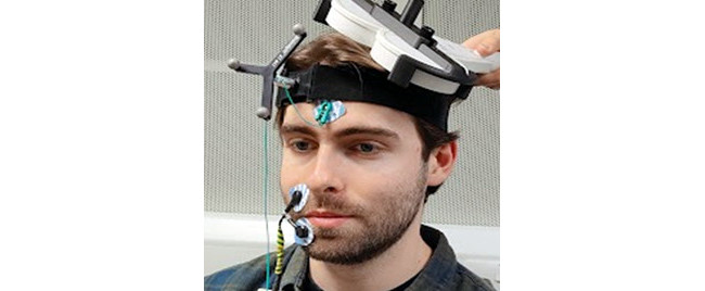
Electrode placement on mouth and forehead
Using this EMG method, we can measure something called a Motor-Evoked Potential (MEP). An MEP is the contraction of a muscle which has been elicited by us externally – by using the TMS equipment. The TMS pulse induces an action potential in the lip region of the motor cortex, and that leads to muscle in the lip to contract – this contraction is what we measure through the MEP. An example of what a standard MEP looks like as recorded by EMG can be seen below. An increased MEP (i.e., a larger amplitude of the wave) indicates that the motor system for the muscle of interest is in a state of increased excitability or lowered threshold (Watkins & Paus, 2004). If an MEP is larger during one task than anther, the neural region being stimulated is likely to be more involved in the task with the greater MEP. By comparing these MEPs across participants and conditions, we can better understand how and when the motor system is involved in our listening task.
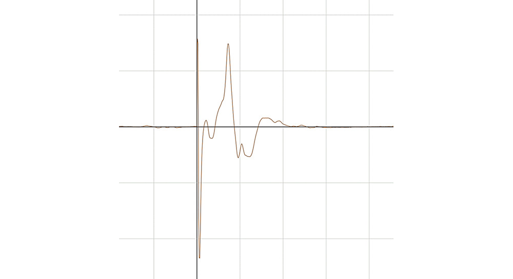
Motor evoked potential (MEP) recorded from lips
The HS-PrediCT team are now underway piloting experiments with this TMS and neuronavigation equipment. We plan to return to you with more blog posts updating you on how piloting has gone, and what tips and tricks for TMS and EMG we’ve learned along the way.
References
Lerner, A. J., Wassermann, E. M., & Tamir, D. I. (2019). Seizures from transcranial magnetic stimulation 2012–2016: results of a survey of active laboratories and clinics. Clinical Neurophysiology, 130(8), 1409-1416. https://doi.org/10.1016/j.clinph.2019.03.016
Rossi, S., Antal, A., Bestmann, S., Bikson, M., Brewer, C., Brockmöller, J., ... & Hallett, M. (2021). Safety and recommendations for TMS use in healthy subjects and patient populations, with updates on training, ethical and regulatory issues: Expert Guidelines. Clinical Neurophysiology, 132(1), 269-306. https://doi.org/10.1016/j.clinph.2020.10.003
Rossi, S., Hallett, M., Rossini, P. M., Pascual-Leone, A., & Safety of TMS Consensus Group. (2009). Safety, ethical considerations, and application guidelines for the use of transcranial magnetic stimulation in clinical practice and research. Clinical neurophysiology, 120(12), 2008-2039. https://doi.org/10.1016/j.clinph.2009.08.016
Wilson, S. M., Saygin, A. P., Sereno, M. I., & Iacoboni, M. (2004). Listening to speech activates motor areas involved in speech production. Nature neuroscience, 7(7), 701-702. https://doi.org/10.1038/nn1263
Fadiga, L., Craighero, L., Buccino, G., & Rizzolatti, G. (2002). Speech listening specifically modulates the excitability of tongue muscles: a TMS study. European journal of Neuroscience, 15(2), 399-402. https://doi.org/10.1046/j.0953-816x.2001.01874.x
Möttönen, R., & Watkins, K. E. (2009). Motor representations of articulators contribute to categorical perception of speech sounds. Journal of Neuroscience, 29(31), 9819-9825. https://doi.org/10.1523/JNEUROSCI.6018-08.2009
Bartoli, E., D'Ausilio, A., Berry, J., Badino, L., Bever, T., & Fadiga, L. (2015). Listener–speaker perceived distance predicts the degree of motor contribution to speech perception. Cerebral Cortex, 25(2), 281-288. https://doi.org/10.1093/cercor/bht257
Murakami, T., Restle, J., & Ziemann, U. (2011). Observation-execution matching and action inhibition in human primary motor cortex during viewing of speech-related lip movements or listening to speech. Neuropsychologia, 49(7), 2045-2054. https://doi.org/10.1016/j.neuropsychologia.2011.03.034
Panouillères, M. T., & Möttönen, R. (2018). Decline of auditory-motor speech processing in older adults with hearing loss. Neurobiology of Aging, 72, 89-97. https://doi.org/10.1016/j.neurobiolaging.2018.07.013
Wendt, D., Kollmeier, B., & Brand, T. (2015). How hearing impairment affects sentence comprehension: Using eye fixations to investigate the duration of speech processing. Trends in Hearing, 19, 2331216515584149. https://doi.org/10.1177/2331216515584149