Proposed national facility for 11.7T human MRI scanning
High magnetic field strength offers great benefits for magnetic resonance imaging, providing improved spatial and temporal resolution, greater sensitivity to physiological changes and access to new contrasts.
With new advances in magnet design and construction, and the recent development of novel, enabling technologies, there is an opportunity for the UK to develop and exploit a very high field MRI capability, matching planned developments in France, Germany, Korea and the Netherlands. Below, we outline the case for an 11.7T scanner to be operated as a national facility for the UK.
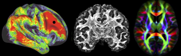
The new scanner will allow us to push the boundaries in resolution for imaging function/ microstructure/connectivity in the brain and other organs, beyond the limits of what is currently feasible, exampled in the Figure, for understanding with higher specificity health and disease.
Background
Magnetic resonance imaging (MRI) and magnetic resonance spectroscopy (MRS) are key tools in medical research, providing information about anatomy, physiology and biochemistry in the human body non-invasively. MRI and MRS underpin a broad range of clinical and neuroscience-focused research programmes in the UK.
They have enabled a vast range of experimental medicine studies into the origins of disease and mechanisms and efficacy of pharmaceuticals and other treatments. High magnetic field strength offers great benefits for MRI, providing improved spatial and temporal resolution, greater sensitivity to physiological changes - particularly for functional brain imaging - and access to new contrasts, such as those based on chemical exchange saturation transfer and magnetic susceptibility. High field also delivers greater spectral dispersion and enhanced sensitivity for MRS based on measurement of signals from 1H nuclei in metabolites. It also opens up new possibilities for metabolic and kinetic studies based on measurement of the signals from other nuclear species, including 2H, 13C, 17O, 31P, 23Na, 39K, 35Cl and even 7Li.
The new 11.7T scanner would be operated as a national facility to capitalize on the UK’s strength in medical imaging.
Realising the benefits of high field requires a programme of work to overcome the significant challenges involved in operating at higher magnetic resonance frequencies. This demands new approaches to the physics of MRI and MRS and the development of novel technologies. In recent years significant scientific and technological progress has been made in addressing some of these issues at 7T and it is now time to extend this work to allow us to establish 11.7T as an important new resource for biomedical research.
Proposal
We propose the establishment of the UK’s first 11.7T MRI scanner for imaging and spectroscopy. This scanner would be operated in Nottingham as a national facility, providing a platform for world-leading neuroscience-focused and clinical studies. In particular, an 11.7T facility would:
- Allow fMRI data to be routinely acquired at 500 μm isotropic resolution, facilitating the characterisation of the laminar and columnar architecture of the human brain over extended cortical regions
- Provide anatomical brain images with sub-100 μm resolution which will facilitate the identification of changes in brain architecture and microstructure in neurodegenerative diseases (including Alzheimer’s, Parkinson’s and Huntington’s disease), and in a range of neurodevelopmental disorders (including autism and schizophrenia) opening a new window on the gene-brain interactions that underpin brain health
- Allow dynamic, spatially resolved and quantitative measurement of brain, liver and muscle metabolism and bioenergetics using 1H, 31P and 13C MRS with measurement volumes that are 10 times smaller than is possible at 3T, opening up new ways to study changes in biochemistry and neurochemistry in a range of disorders
- Provide exquisite functional and biochemical interactions to study interacting effects between organs for instance in conditions such as diabetes and the complex effects of multi-morbidity
- Form a strong focus for the development of new MR technology and image processing methodology – which experience shows will also yield benefits for MRI/S at lower field strengths
System
An 11.7T magnet with a 830-mm bore, which could be manufactured from niobium-titanium superconducting wire cooled to 2.2 K. The magnet will require passive shielding (~500 tonnes of iron).
Scanner electronics would be provided by one of the major scanner manufacturers who would take responsibility for system integration.
RF coils would be developed in-house and in collaboration with third party manufacturers.
Management
We expect that the new scanner will be operated as a national facility, and housed in an extension of the Sir Peter Mansfield Imaging Centre building at the University of Nottingham. Through its establishment of a Beacon of Excellence in Precision Imaging, the University of Nottingham aims to develop the next generation of biomedical imaging systems that will underpin the future exploitation of imaging in precision medicine across the world.
This new facility will also provide research space for visiting researchers, as well as facilities for patients/experimental subjects. A steering committee with representation from leading UK centres will be established to oversee the development of the scanner and manage access to the 11.7T facility. We also have very good links with the European, Asian and US sites that are already operating human MR scanners at 9.4 and 10.5T (Tübingen, Maastricht, Jülich, Minnesota and Chicago), and those planning to work at 11.7T (NIH, Neurospin – CEA-Saclay, Gachon University), as well as the German and Dutch consortia who are planning the development of 14T systems, and can call on a wide range of international expertise to support a UK effort in UHF MRI/S.
Community Statement of Need (June 19)
Preliminary IAC Application (April 20)
