Computed Tomography (MicroCT) for Other Applications
These 3D visualisations were created from MicroCT datasets. Through advanced image analysis techniques, it is possible to separate the different phases of materials within a sample and false colour them for clarity.
Food Technology

Aerated chocolate permitting analysis of 3D porosity and bubble size distribution.
Biomaterials
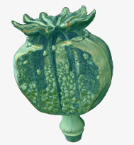
Poppy seed pod allowing automatic assessment of inclusions and particles
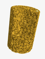
Engineering
Metal wires in a cement core allowing evaluation of strength characteristics
Pharmaceutical
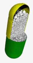
Paracetamol capsule showing visualisation of particle packing density
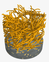
Electronics

A USB stick allowing detailed component analysis and defect detection
Geosciences
Sandstone scanned to reveal changes in morphology following fluid flow