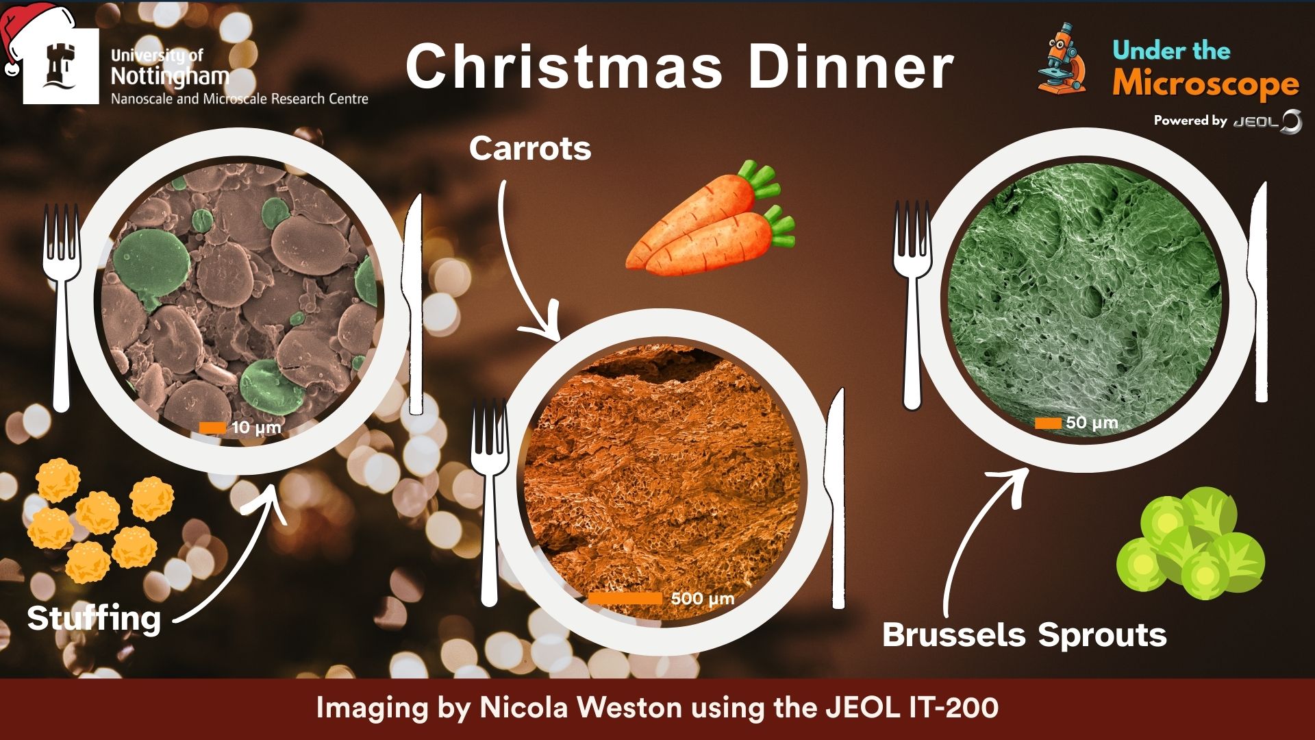
December 2025 - Christmas Dinner
Fair to say that when it comes to the nation's favourite annual meal, we're often practising speed and not attention! But have you ever wondered how the components of a stuffing mix come together? What the structure of your carrot is that gives it the perfect crunch? Or even just what on earth is a brussels sprout really about??
The December edition of Under the Microscope powered by JEOL gives us all a sneaky peek at these exact things. Fascinating!! Thanks to Nikki Weston for imaging on the JEOL IT-200 and to Sally Schofield and many curious school children across Nottinghamshire for suggesting!
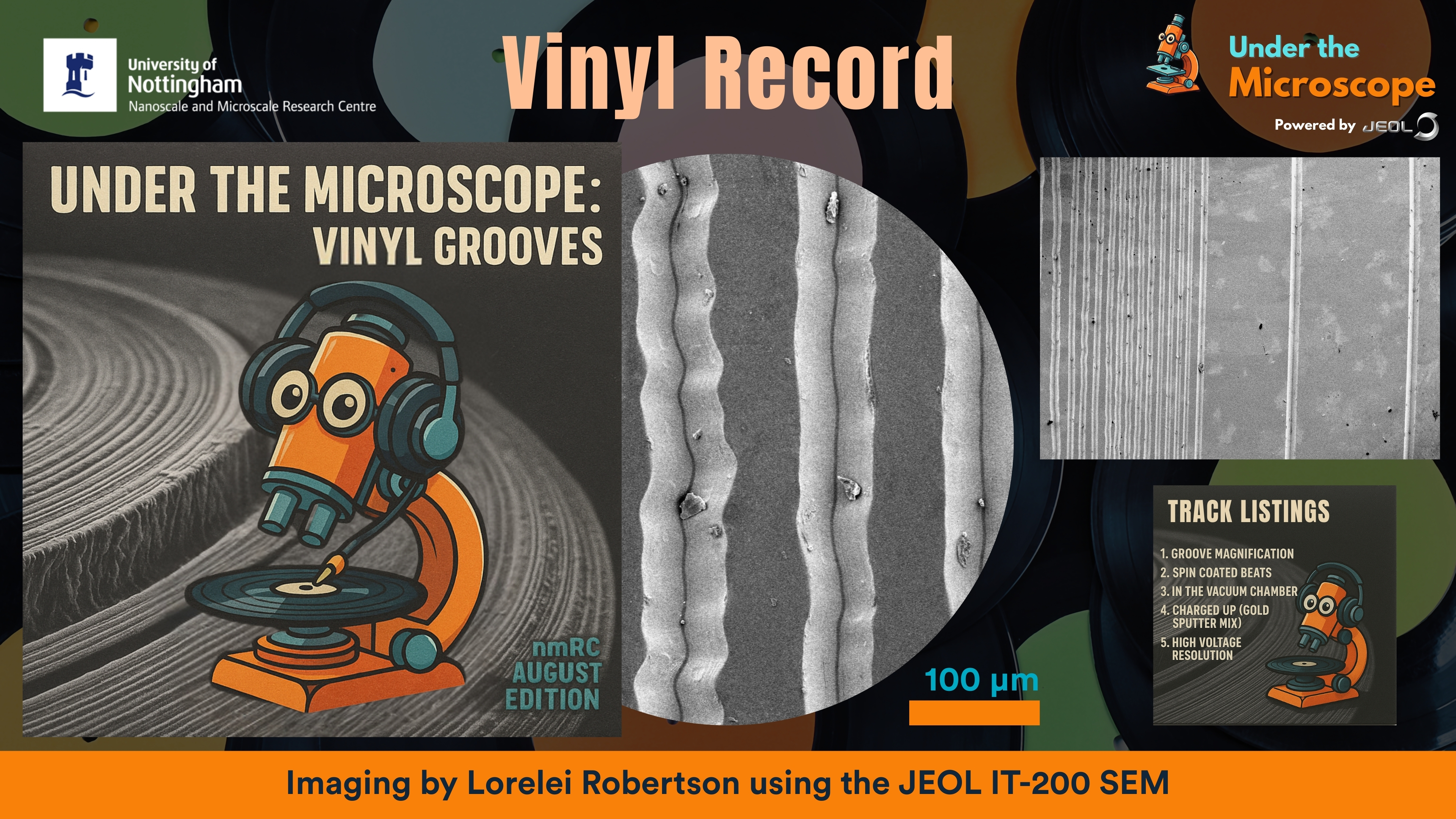
August 2025 - Vinyl record
This August, we put a spin on our Under the Microscope powered by JEOL series by imaging a vinyl record with our IT-200 scanning electron microscope (SEM) - thanks to Dallas Campbell for the suggestion!
What looks like smooth black plastic to the naked eye transforms under the SEM into a stunning landscape of intricate grooves — tiny canyons that carry the music we love.
To celebrate, we created a mock vinyl sleeve cover featuring our mascot, Scopey , DJ-ing on the record itself.
Why This Is Cool:
SEM lets us see the incredible detail and precision of vinyl grooves at the microscale.
Those wavy patterns are the very soundwaves etched into the record!
It’s a fun reminder of how science meets art and technology in everyday objects.
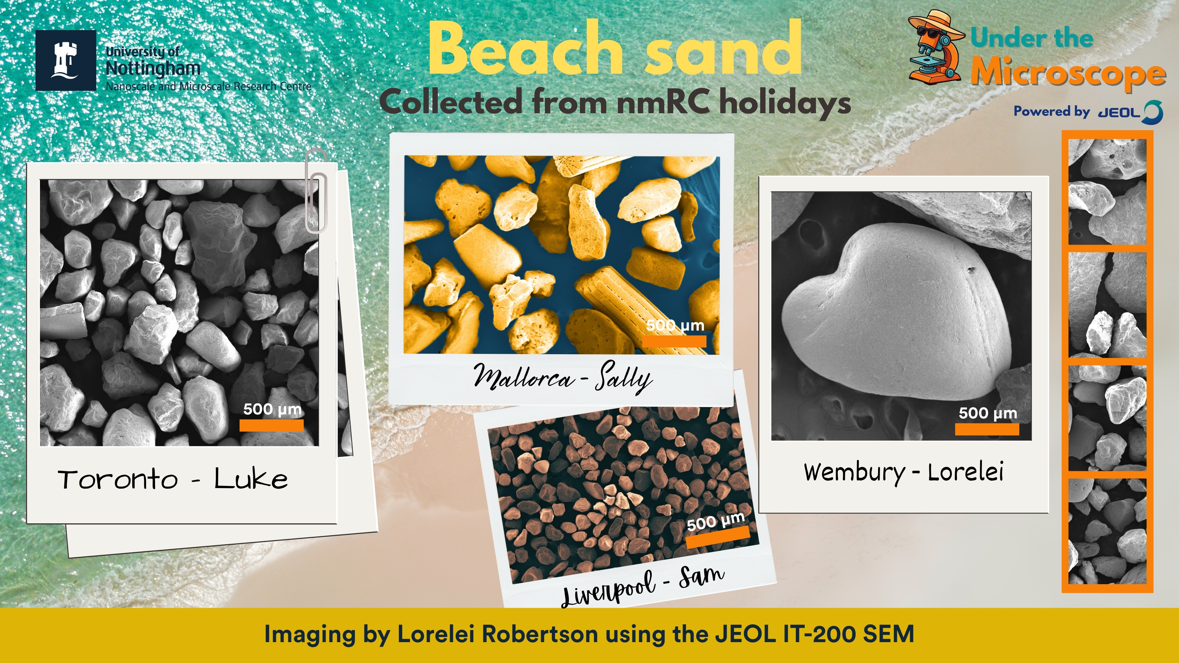
This month at the nmRC, "our job is actually just, you know, Beach!"
We’re bringing a bit of the sunny season to the lab with our Under the Microscope powered by JEOL initiative! Inspired by staff holidays in the sun, we’ve been imaging sand samples from around the world – each grain a tiny postcard from nature.
The idea came from Greg Corbett, with beautiful imaging by Lorelei Robertson on our JEOL IT-200 SEM.
Featured locations and collectors:
Wembury, UK – Lorelei
Mallorca, Spain – Sally
Liverpool, UK – Sam
Toronto, Canada – Luke
From volcanic fragments to coral remnants, the SEM reveals a hidden universe beneath our feet – or flip-flops!
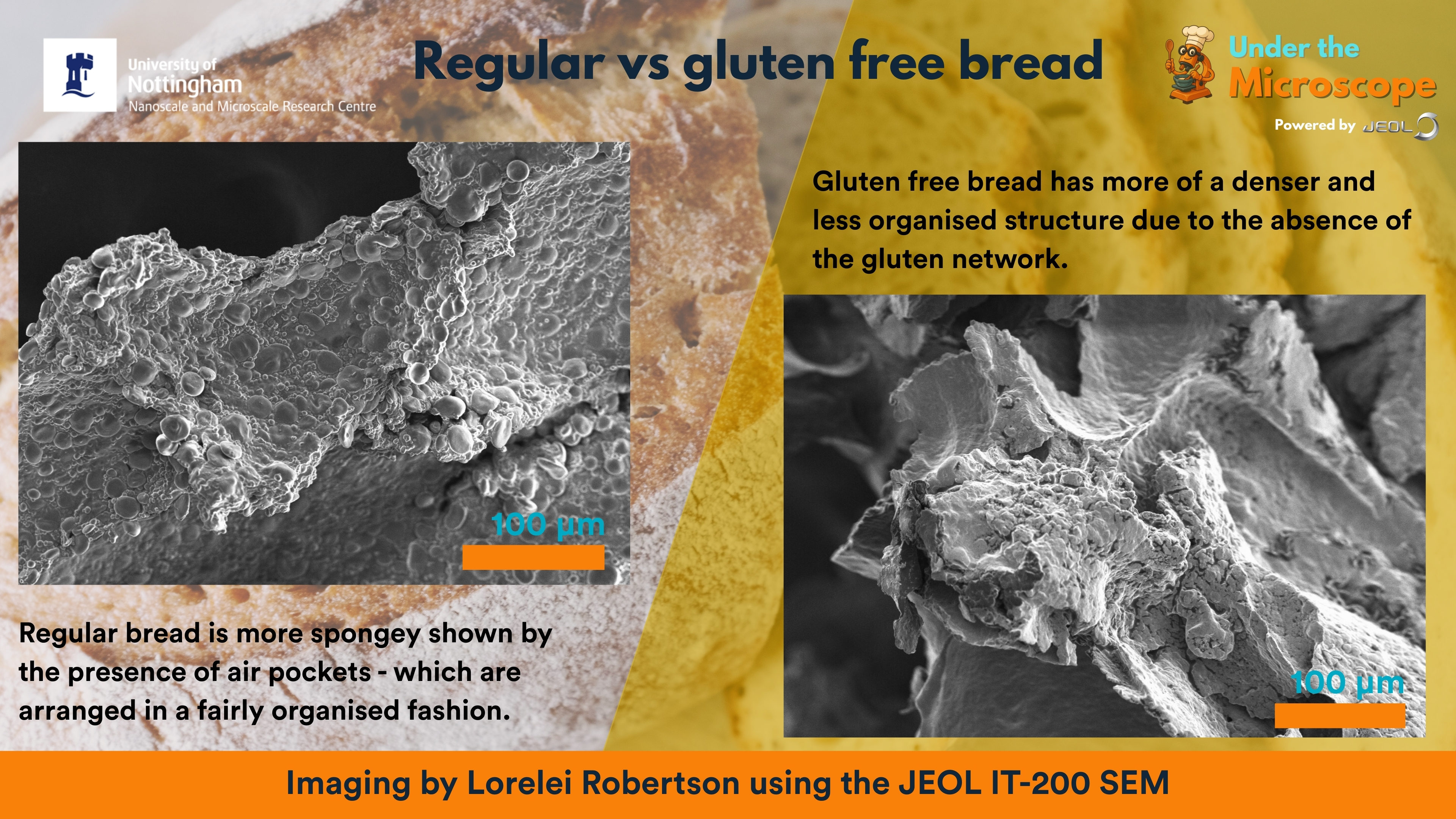
April 2025 - Bread vs Gluten Free Bread
The best thing since sliced bread... at high magnification! The images are loaf at first sight!
Thanks to Susanne, last month’s winning suggestion, we dove into the microscopic world of bread vs gluten-free bread—and the results are fascinating!
Using our JEOL IT-200 Scanning Electron Microscope, Lorelei Robertson (Electron Microscope Technician) captured the intricate textures and structures that make these everyday loaves so different at the nanoscale.
From gluten’s stretchy protein networks of air pockets to the gluten free's more denser structure, this month’s images reveal just how much is happening beneath the crust.
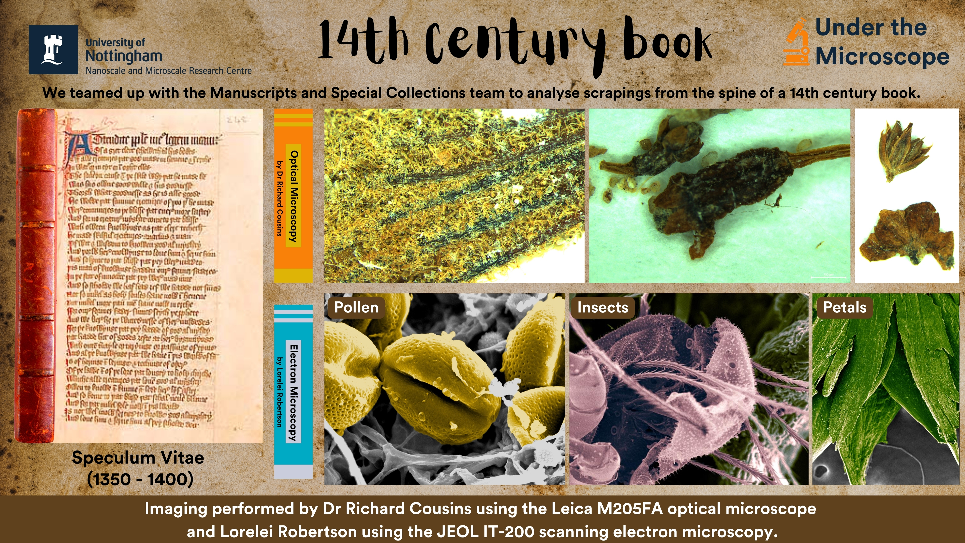
February 2025 - 14th Century Book
February’s Under the Microscope winning suggestion comes from our colleagues at the Manuscripts and Special Collections team (Dr Charlotte May and Robert Pearce) and what a fantastic choice it was!
At the nmRC, we love uncovering the hidden stories within historical objects. Our latest collaboration has taken us deep into the spine of a 14th-century manuscript, Speculum Vitae (Mirror of Life), revealing a microscopic world preserved for centuries.
Using optical and electron microscopy, we examined material collected from the book’s spine—fragments of plant matter, pollen grains, flower petals, and even tiny micro-creatures. This nanoworld, frozen in time, offers us a glimpse into the past, providing clues about how this manuscript was used and cared for over the centuries. Imaging by Lorelei Robertson and Dr Richard Cousins using a Leica optical microscope and a JEOL IT-200 scanning electron microscope.
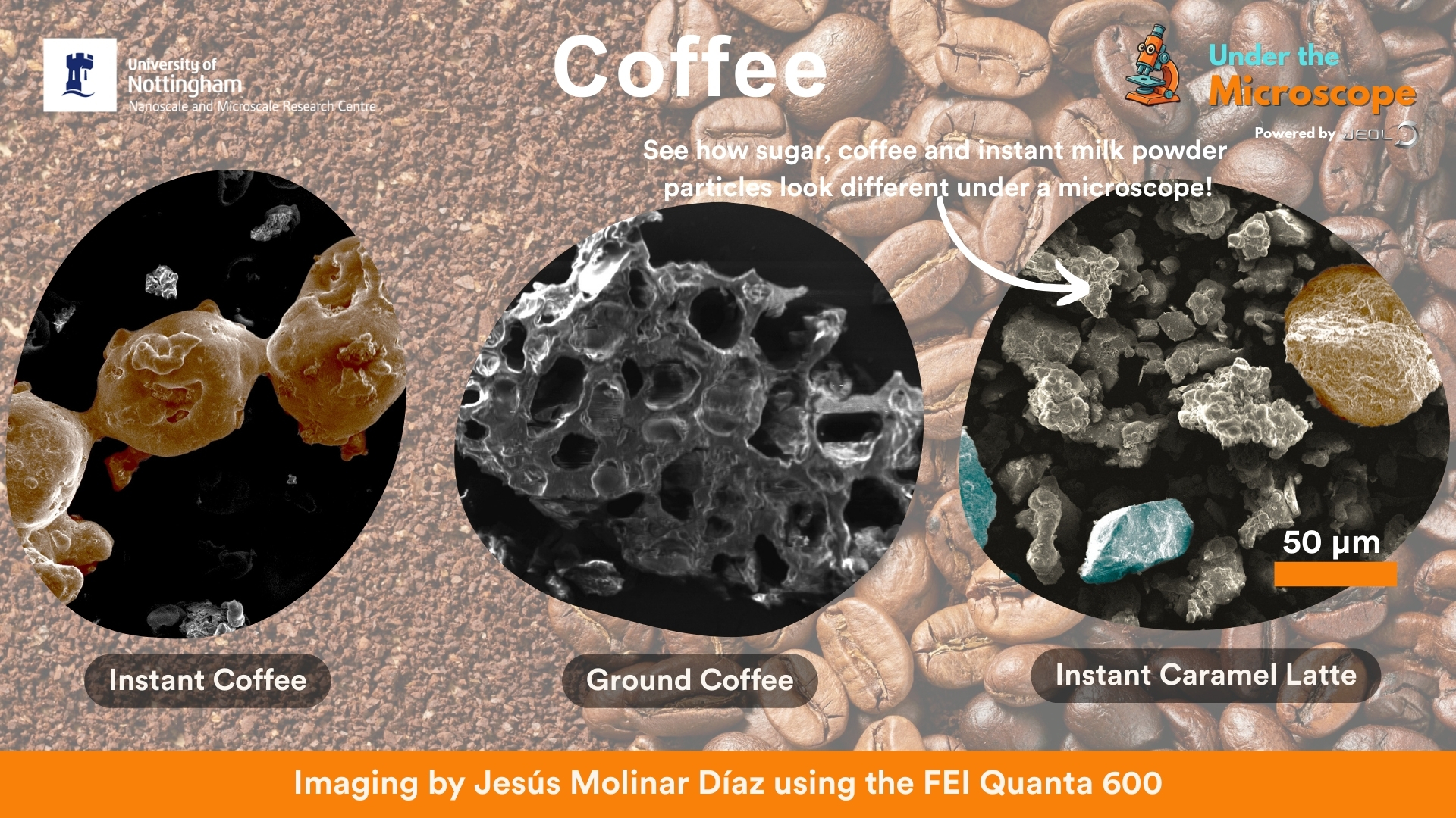
This November we're tackling a popular suggestion for the Under the Microscope powered by JEOL, Coffee. Thanks to: Rob, Ian Siara and Sam Kilgour for the suggestion.
Jesús imaged three different types of coffee using our Quanta 600 SEM: Instant coffee, Ground coffee and instant caramel latte. The images showing a difference in the microstructure between instant and ground coffee, as well as being able to see the different constituents that make up an instant Latte: Coffee, milk powder and sugar.
September 2025 - Tardigrade
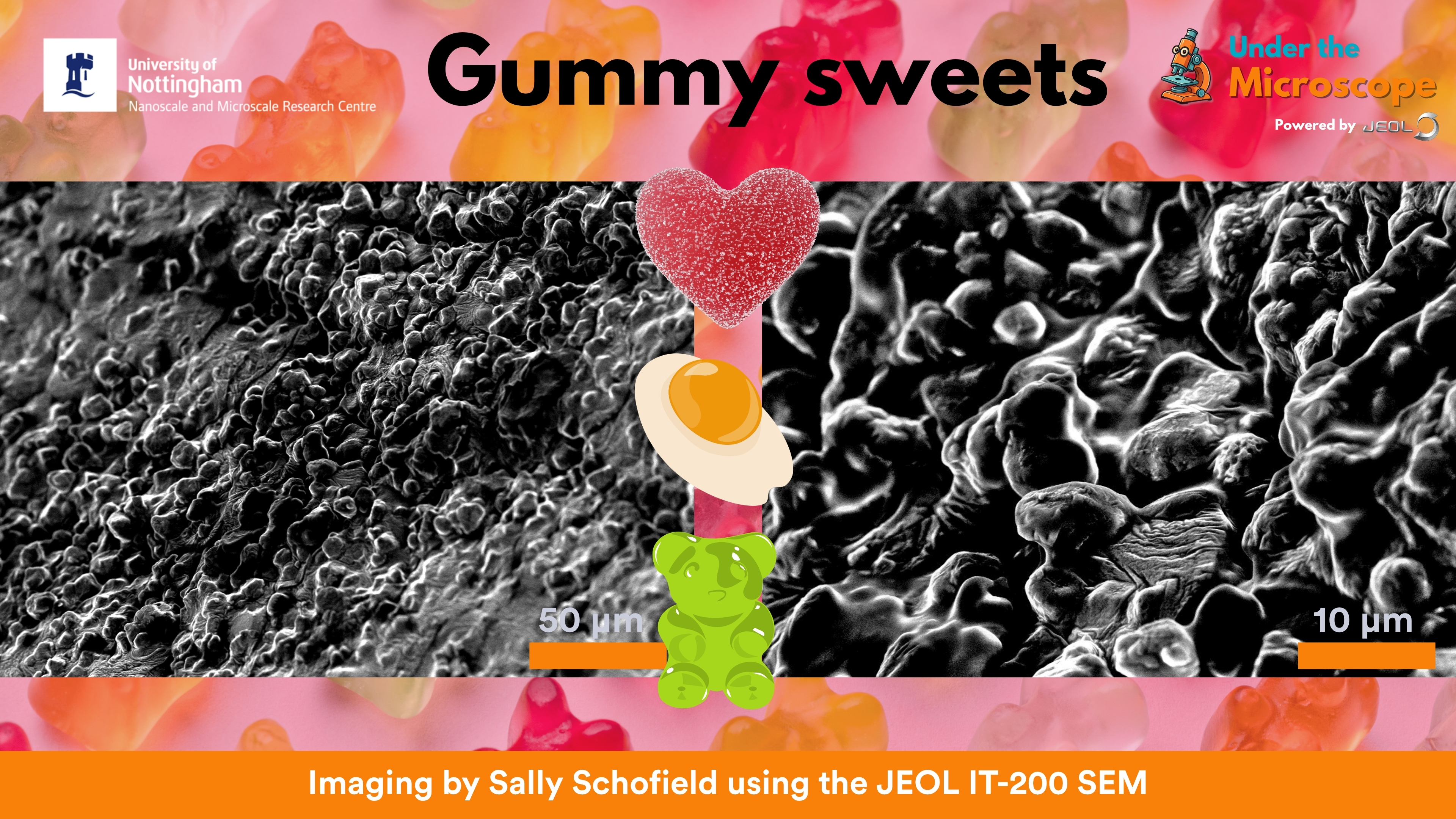
Our latest Under the Microscope powered by JEOL winner is… Gummy Sweets! (suggested by Julia Siara)
Who knew your favourite chewy snack could look so fascinating up close?
Our JEOL IT-200 SEM (operated by Sally Schofield) captured the hidden world inside these sugary treats – proving that science can be as sweet as candy!
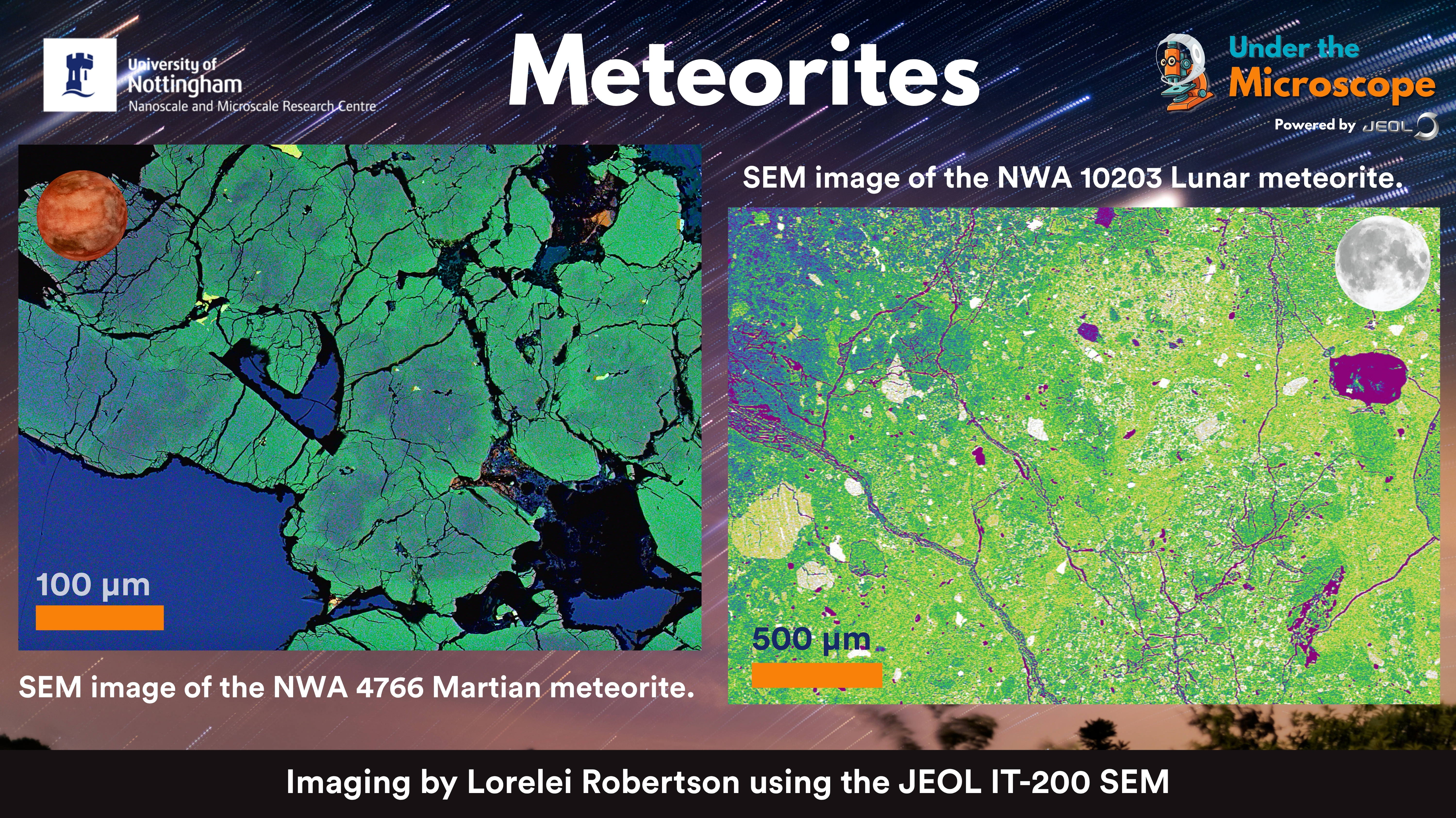
May's winning suggestion for the nmRC’s Under the Microscope powered by JEOL was meteorites, a stellar idea from "Marie"!
We put fragments of Lunar and Martian meteorites under the Scanning Electron Microscope (SEM) to reveal their extraordinary microstructures — a rare glimpse into the geology of other worlds.
These samples aren’t just space rocks — they’re fragments of the Moon and Mars that travelled millions of miles to end up here on Earth. The SEM images reveal stunning details of their mineral composition, shaped by cosmic forces far beyond our planet.
Imaging was performed by Lorelei Robertson using the JEOL IT-200 SEM.
March 2025 - Mosasaur Tooth
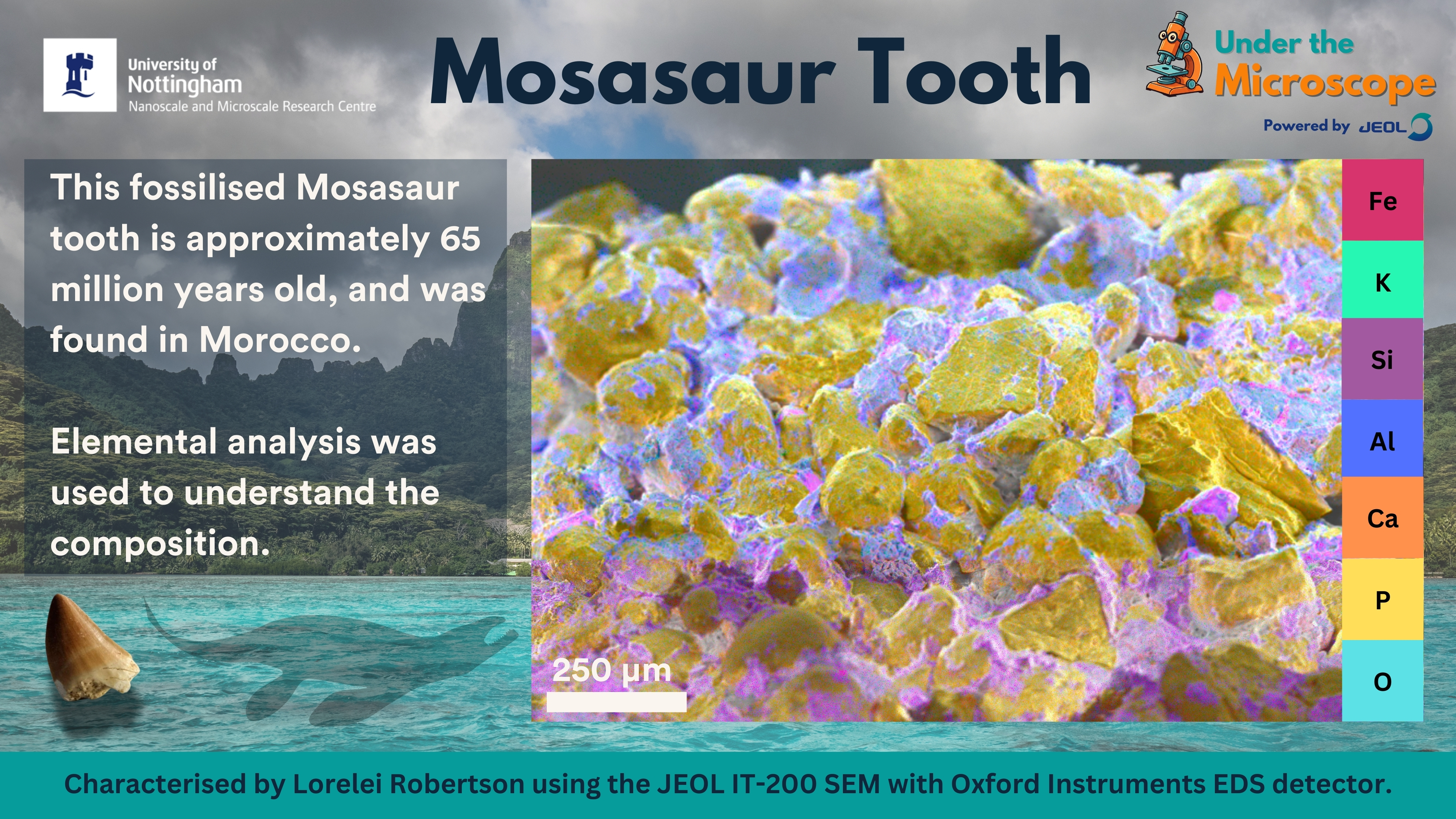
March's Under the Microscope takes us back 65 million years to the age of marine reptiles! Meet the mosasaur, a formidable apex predator whose fossilised tooth we’ve imaged using Scanning Electron Microscopy with Energy-Dispersive X-ray Spectroscopy (SEM-EDS).
These high-resolution images reveal the microscopic structure and elemental composition of the tooth, offering insights into its preservation and the ancient marine environment it came from.
⭐ Suggestor wished to remain anonymous but a huge thank you for your great idea!
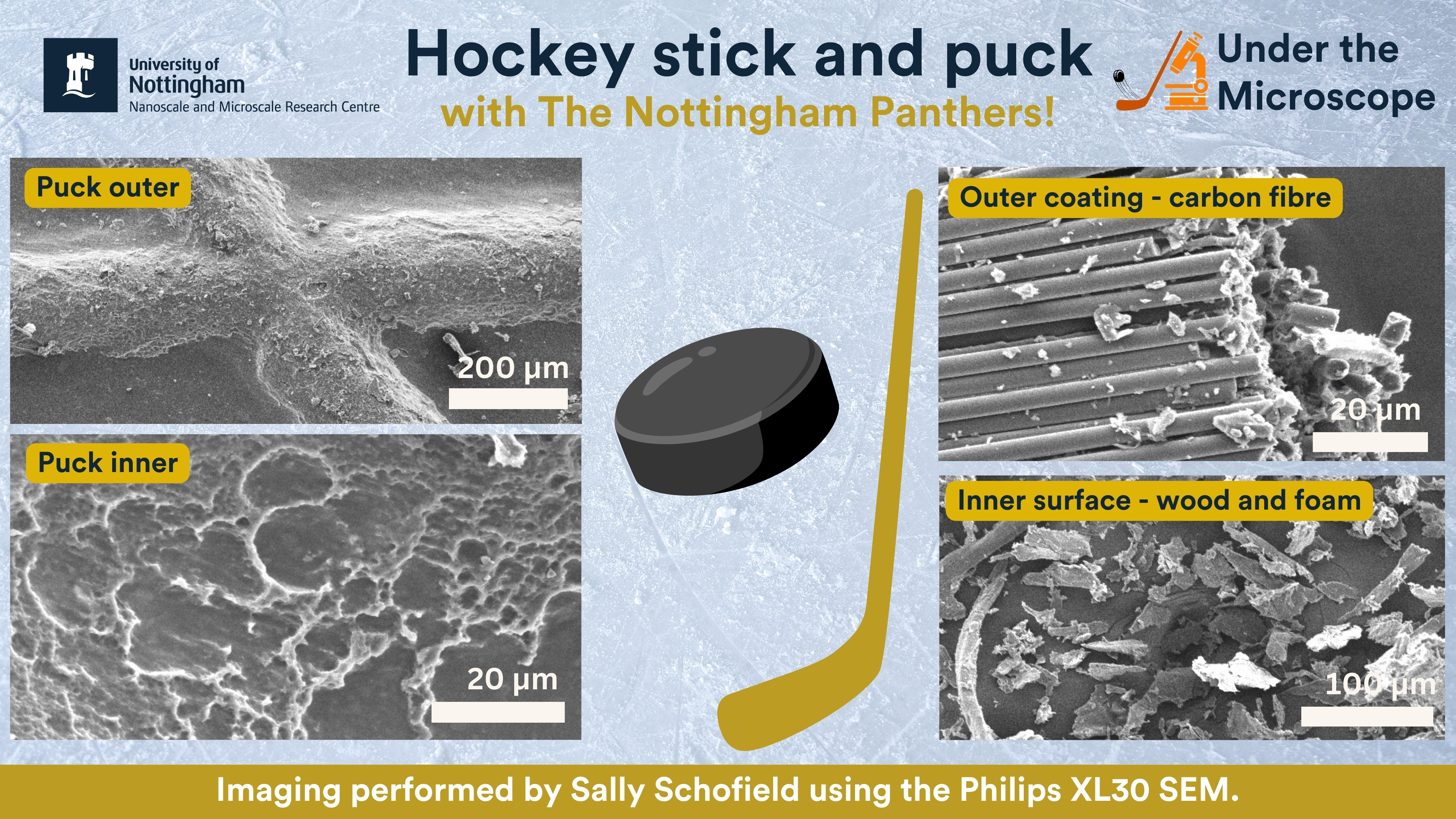
January 2025 - Ice Hockey Puck and Stick
Our first Under the Microscope of 2025 takes a cool look at an ice hockey puck and stick, generously donated by the Nottingham Panthers!
Using the Philips XL30 SEM, our laboratory support technician (and Panthers superfan) Sally Schofield captured stunning electron microscope images revealing the hidden details of these essential pieces of hockey gear. The puck's tough rubber exterior was clearly visible, showcasing its resilience on the ice. Meanwhile, the stick’s wood and foam interior and carbon fibre coating were revealed in intricate detail—highlighting the balance of strength and flexibility needed for that perfect slapshot!