Dynamic Nuclear Polarisation: a new MR technology for metabolic studies in health and diseas
Nuclear Magnetic Resonance (NMR) spectroscopy and Magnetic Resonance Imaging (MRI) provide non-invasive insight into structure, dynamics, and metabolism across the physical, biological, and medical sciences. In exercise physiology, these techniques are indispensable for probing skeletal muscle bioenergetics in vivo, yet their limited sensitivity restricts the detection of rapid metabolic fluxes and low-abundance metabolites. In the brain, spatial mapping of these metabolites is critical for identifying metabolic changes associated with neuronal activity or disease, but similar sensitivity limitations persist.
By using Dynamic Nuclear Polarisation (DNP) to hyperpolarise 13C-labelled pyruvate, nuclear spin polarisation—and thus signal sensitivity—can be enhanced by several orders of magnitude (10,000-fold). This sensitivity gain enables real-time, non-invasive measurements and mapping of pyruvate metabolism in vivo, including its conversion to lactate, alanine, and bicarbonate in the whole body and the brain.
DNP applications in human skeletal muscle studies
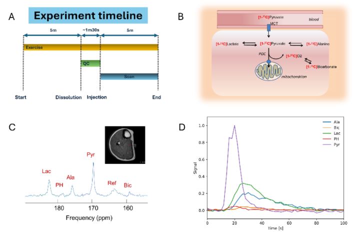
Hyperpolarised 13C-MR spectroscopy detects muscle pyruvate during steady-state exercise in participants. (A) 5 min after exercise, dissolution is started and followed by ~ 90s of quality control check before injection into the volunteer. Then the MR scan starts and is acquired over 5 min (flip angle 49±7°, bandwidth 5 kHz, TR 2 s, 2048 complex points, duration 300 s). (B) Labelling of cell metabolites from hyperpolarised [1-13C] pyruvate. (C) Representative mDIXON images and 13C NMR spectra showing muscle volume and 13C pyruvate conversion to lactate (lac), pyruvate hydrate (PH), alanine (ala), reference (ref) and bicarbonate (bic) in participants exercising. The internal reference (ref) was a fiducial containing 8 M 13C urea. (D) Representative time-courses of the normalised signal amplitudes of 13C-labelled metabolites after injection of hyperpolarised [1-13C] pyruvate in one participant during MR in bore exercise
DNP applications in human brain studies
Traditionally, 31P-MRS has been the gold standard for assessing skeletal muscle energetics, providing indirect insight into oxidative metabolism through measurements of phosphocreatine, ATP, and intracellular pH. While powerful for evaluating mitochondrial function and recovery kinetics, it does not directly measure substrate flux. Hyperpolarised 13C-MRS offers a complementary approach by dramatically enhancing sensitivity via dynamic nuclear polarisation of 13C-labelled pyruvate, enabling real-time, non-invasive assessment of pyruvate metabolism in vivo. The detection of 13C-bicarbonate provides a direct marker of pyruvate dehydrogenase flux. Combined 31P- and 13C-MRS enable integrated assessment of muscle bioenergetics and pathway-specific metabolism during exercise, adaptation, and disease
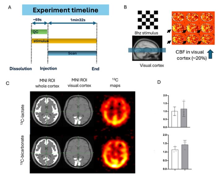
Hyperpolarised 13C-MR spectroscopy detects brain oxidative metabolism during visual activity. (A) Dissolution and stimulus start at the same time, and injection of HP-13C pyruvate (~69s after dissolution) is followed by a spectral-spatial 13C imaging scan (1min32s) performed at 3T using a modified 3D echoplanar sequence with B1 and B0 map correction (flip angles set to 5° for pyruvate, 15° for lactate, and 60° for bicarbonate, with an isotropic resolution of 4.5 cm³; manuscript in preparation). (B) A 8 Hz flashing checkerboard was used as visual stimulus for HP-13C and ASL scans, with 3D T1-weighted images acquired for anatomical co-registration. Basal CBF in the visual cortex increases during stimulus (~20%), confirming increased metabolic demand in response to visual stimuli. (C) T1-weighted images with MNI ROIs and 13C-metabolite maps. (D) Graphs showing a trend towards an increase in the bicarbonate/pyruvate ratio in the visual cortex after stimulation, but not in the lactate/pyruvate ratio.
Hyperpolarised-13C MRI (HP-13C MRI) enables real-time imaging of cerebral metabolism by tracking the conversion of HP-[1-13C] pyruvate to [1-13C] lactate and [13C] bicarbonate. Previous 13C-MR spectroscopy and 13C spectral spatial imaging have reported conflicting results regarding pyruvate metabolism during neuronal activation. Using a highly sensitive 13C spectral-spatial imaging sequence during visual stimulation, we demonstrate consistently elevated 13C-bicarbonate labelling in the visual cortex relative to the rest of the cortex, with no corresponding increase in 13C-lactate. These findings indicate predominantly oxidative metabolism supporting neuronal activation, reveal metabolic heterogeneity in the human brain, and have implications for studying aging and neurological disease, where non-oxidative metabolism becomes more prominent.