Under the Microscope
Have you ever wondered what pollen looks like? Or the scales on a butterfly’s wing? We're giving the public the chance to find out what objects, that usually go unseen, look like with our new ‘Under the Microscope’ initiative.
We are asking for people to suggest objects or materials they would like to see in microscopic detail, as well as explain why e.g. does it have a significant meaning to you? Is it relevant to your local area? etc...
One idea will be selected each month to be imaged by our team using state of the art Scanning Electron Microscopy (SEM) equipment that can create images at the nanoscale. Those images will be sent to the selected entrant and published on this page! To submit your entry please use the link to the right.
What is Scanning Electron Microscopy (SEM)?
Scanning Electron Microscopy (SEM) is a powerful tool used to visualise the micro- and nano-structures of materials. It works by using a beam of negatively charged particles, called electrons, to scan the surface of the material, creating high-resolution images that show details as small as a few nanometres.
To put this into perspective, one nanometre is approximately one million times smaller than the width of a single human hair.
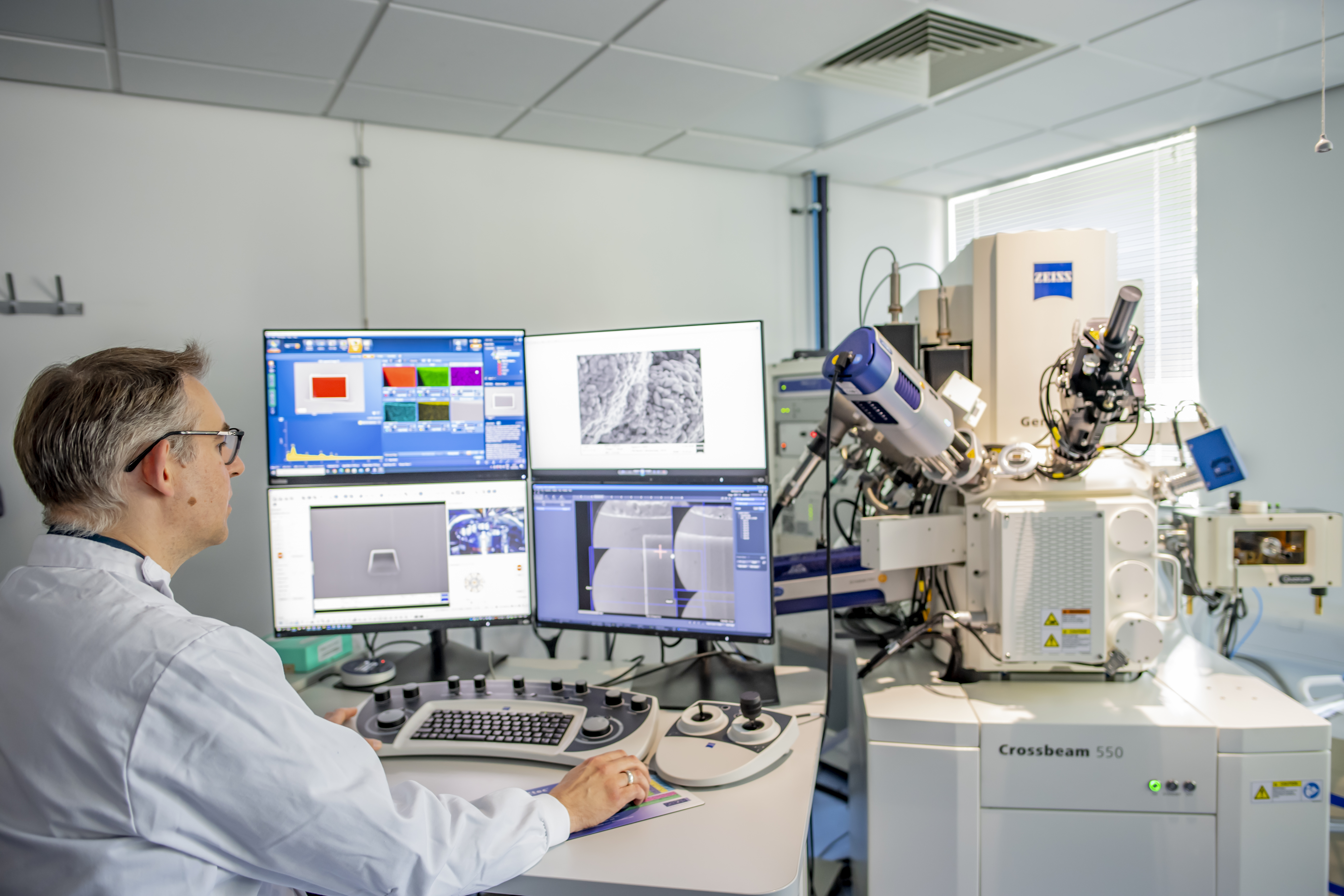
The Zeiss Crossbeam 550 SEM at the nmRC.
Previous Winning Entries
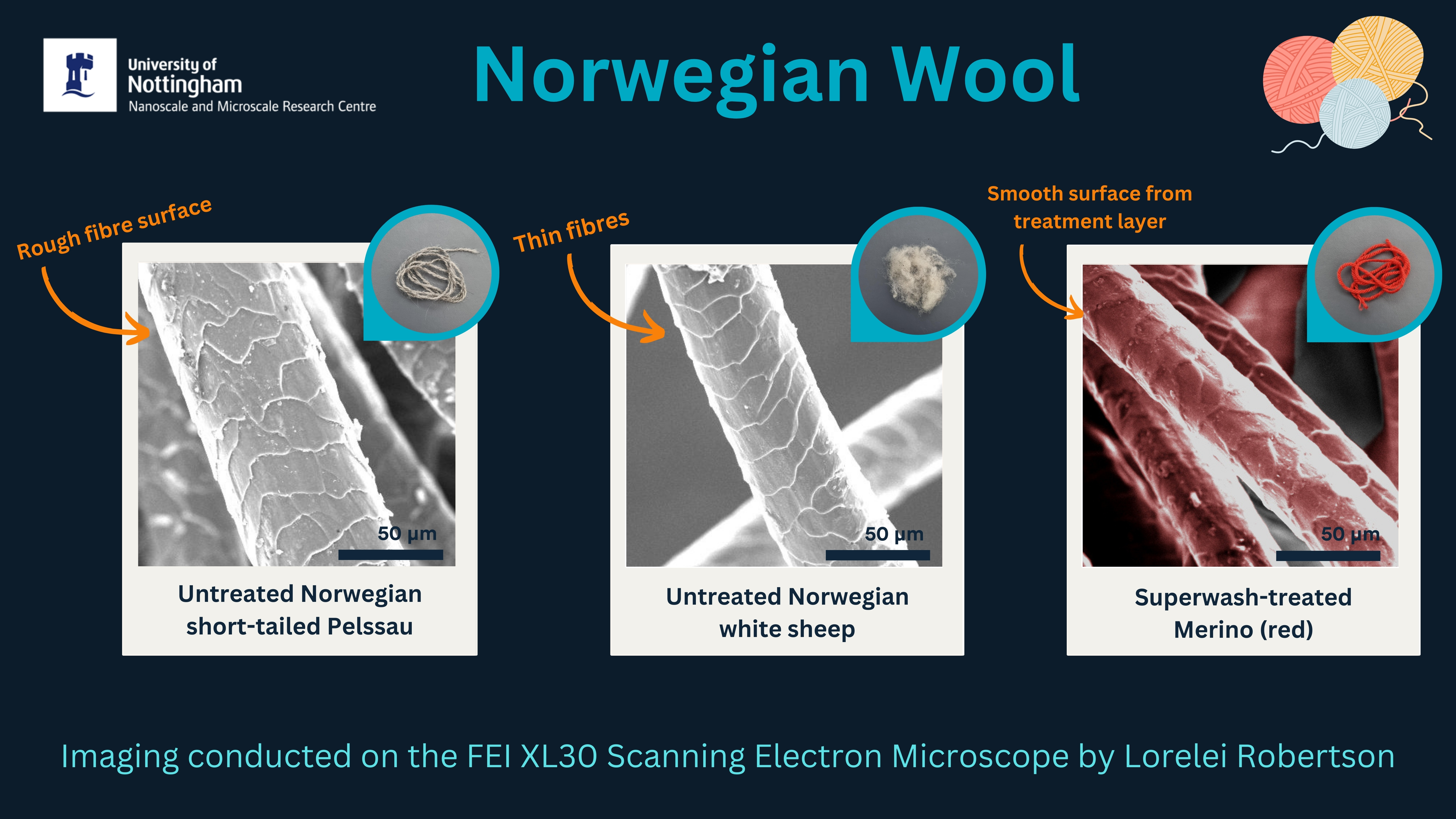
Norwegian Wool - March 2024
It's time for March's Under the Microscope winning suggestion which was...Wool suggested by Kristine Vike. Kristine not only suggested wool but also donated some to us all the way from Norway! For this suggestion, Lorelei Robertson imaged untreated and superwash-treated wools using the Thermo Fisher (FEI) XL30 see if there were any surface differences between the wool threads, and EWE better believe there were!
Snails - February 2024
February's Under the Microscope is here featuring...SNAILS suggested by Lucy Silvey! When it comes to snails, there's only one expert we could turn to: resident snail aficionado Prof. Angus Davison from the School of Life Sciences. He generously provided us with snail shells and even a love dart for imaging! As this marks our 12th monthly suggestion, we're treating you to fluorescence and electron microscopy images of snail shells and love darts. Fluorescence microscopy images were captured by Jordan Kirby using the Leica M205 FA microscope, and electron microscopy images were captured by Lorelei Robertson using the Thermo Fisher (FEI) XL30 SEM. 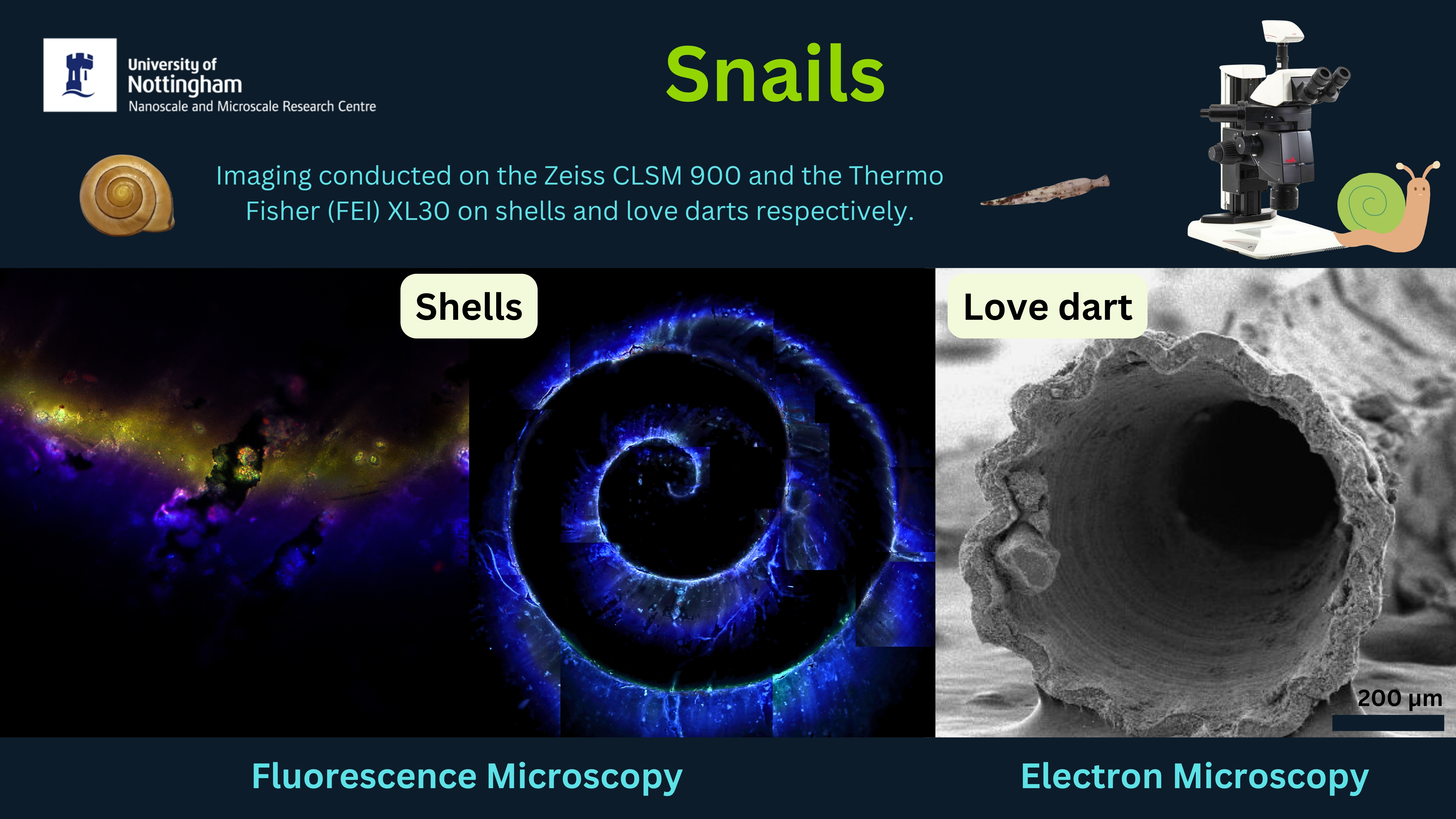
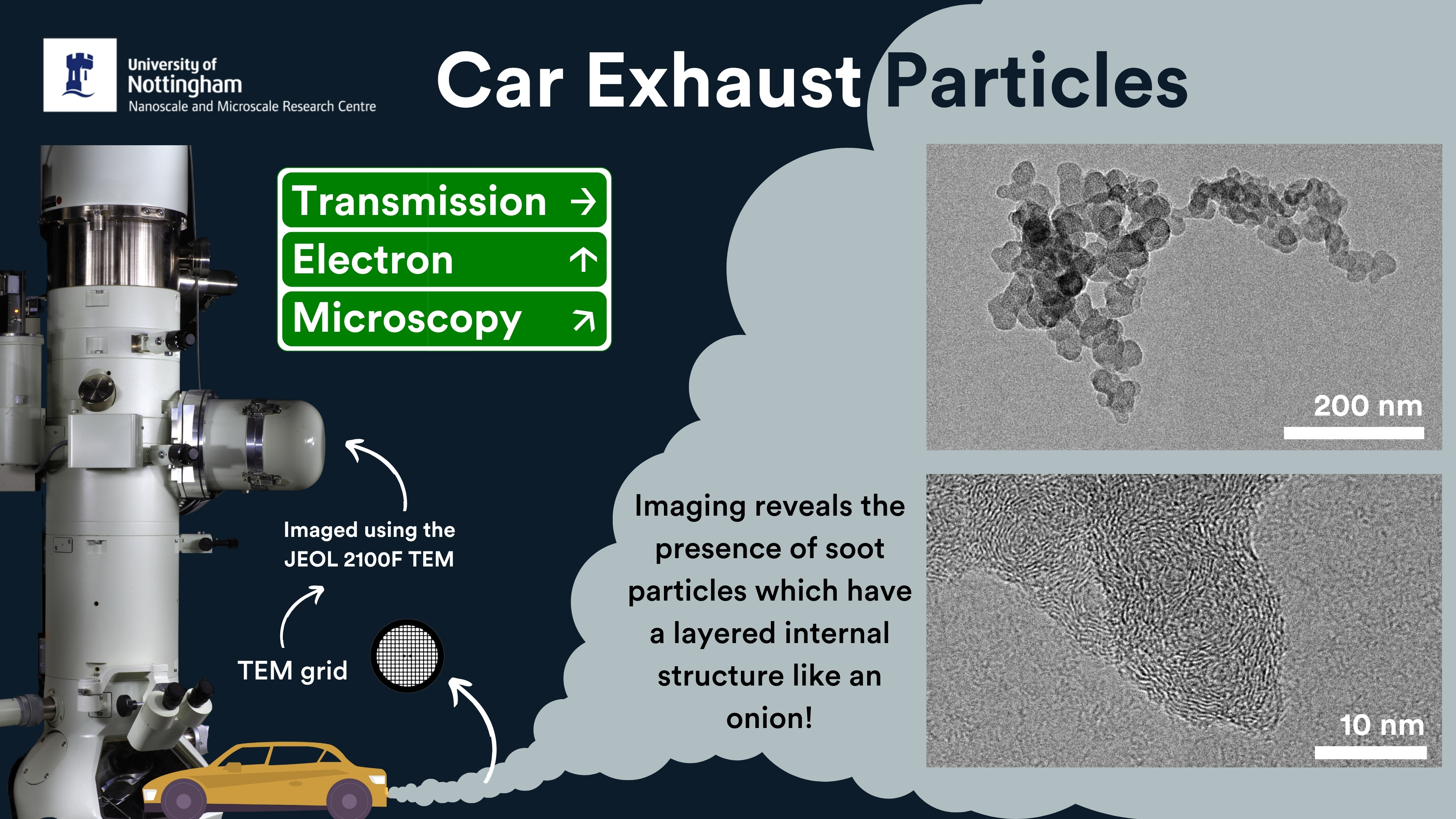
Car Exhaust Particles - January 2024
READY, SET, MAGNIFY! We are kicking off Under the Microscope 2024 with some Transmission Electron Microscopy (TEM) images of car exhaust particles, suggested by Dr Tendai Dube! These TEM images show the presence of soot particles which are the product of incomplete combustion of fuel. The particles, with a diameter ranging from 10 to 20 nm, have a structure consisting of a carbon core surrounded by layers of carbon (resembling nano-onions). Imaging was conducted by Dr Michael Fay on the JEOL 2100F TEM.
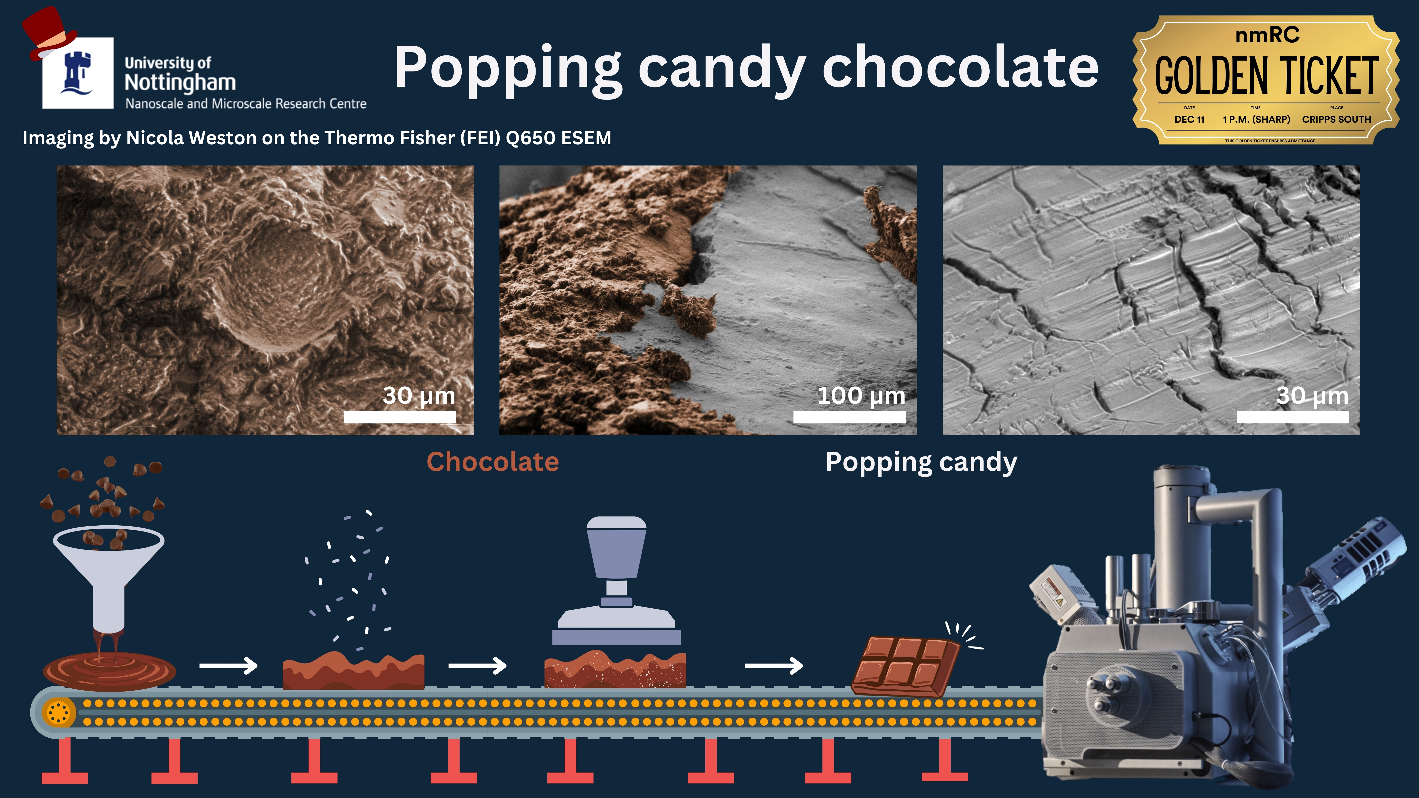
Popping Candy Chocolate - December 2023
This month's Under the Microscope winning suggestion was suggested by our Director Prof. Paul Brown who wanted to celebrate the release of the new film Wonka by imaging some... POPPING CANDY CHOCOLATE! We unwrapped the magic of Willy Wonka's chocolate factory with scanning electron microscopy images of popping candy chocolate that demonstrate a sweet symphony of science and imagination. Imaging conducted by Nicola Weston on the Thermo Fisher (FEI) Q650 SEM.
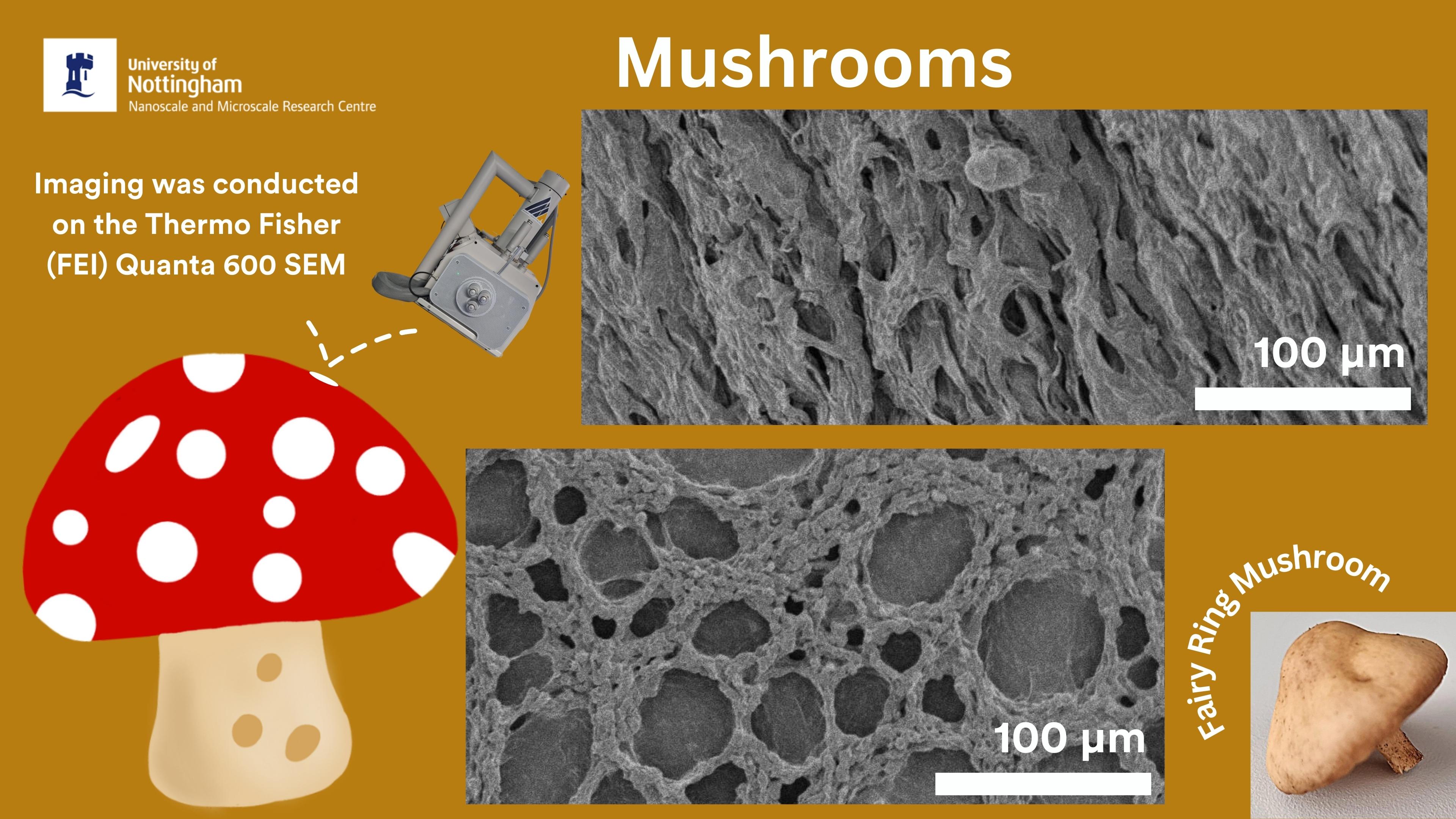
Mushrooms - November 2023
Our Under the Microscope winning suggestion for November was MUSHROOMS which was suggested by Rob Hutchinson and Steph Bowskill. These Fairy Ring mushrooms were found on our campus and we captured so many incredible images of their surface structures because the SPORE the better! SEM Imaging was conducted by Lorelei Robertson using the Thermo Fisher (FEI) Quanta 600 scanning electron microscope.
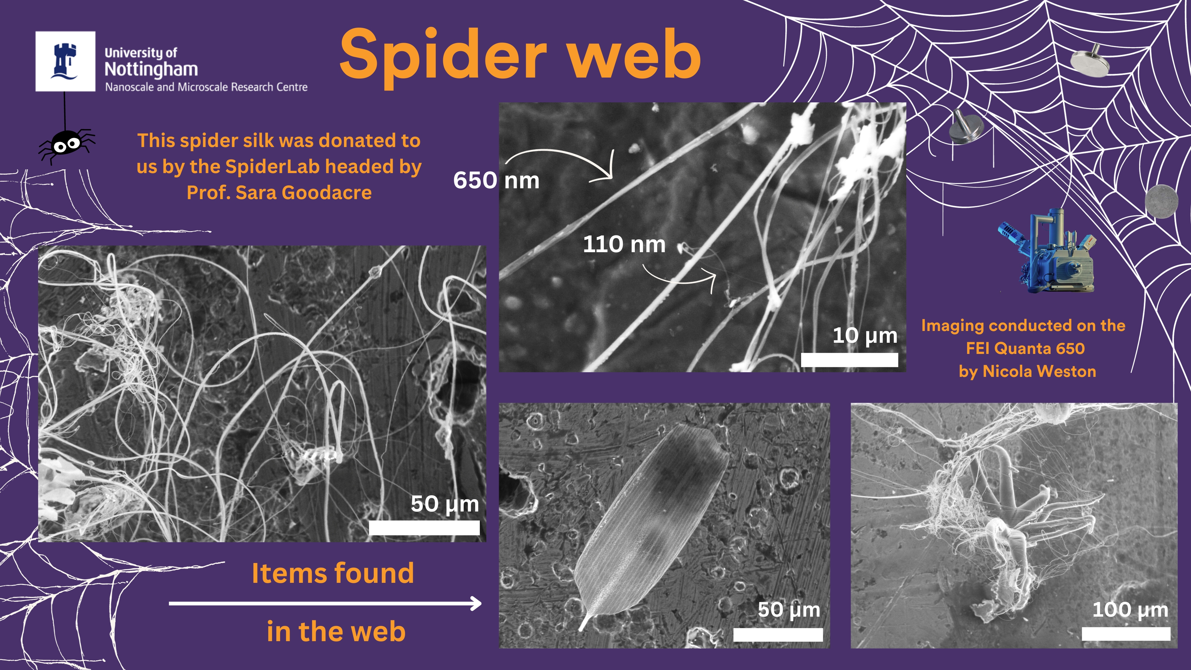
Spider Web - October 2023
We're diving deep into the world of arachnid artistry as this month's Under the Microscope winning suggestion is...Spider Web! The webs were donated to us by Prof. Sara Goodacre of the SpiderLab here at UoN and imaging showed that the thinnest threads were just 110 nm in diameter. We also found an array of different objects that had been caught in the webbing. SEM imaging was conducted by Nicola Weston on the Thermo Fisher (FEI) Quanta 650. Thanks to Reza for the suggestion!
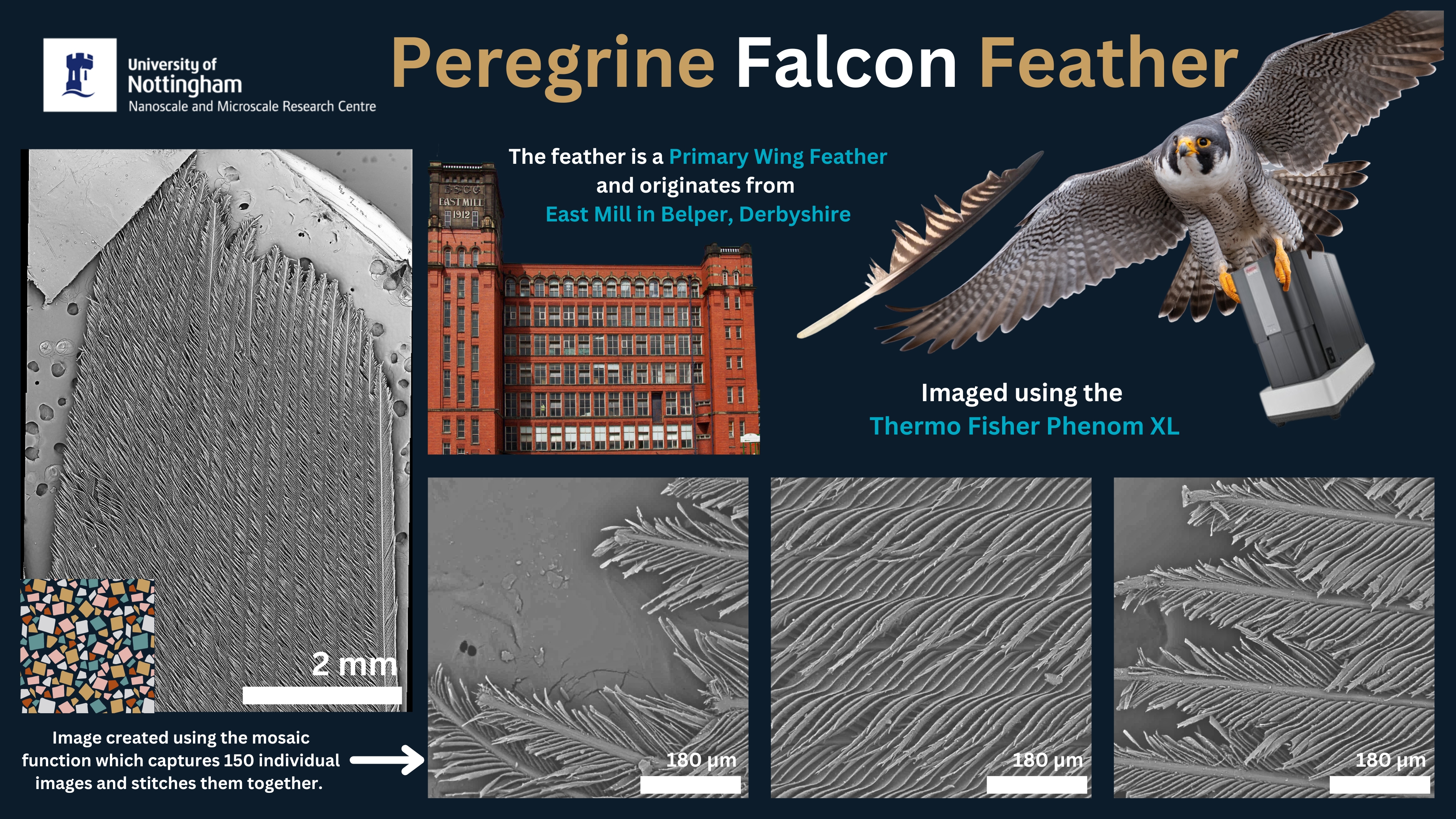
Peregrine Falcon Feather - September 2023
This month's Under the Microscope winning suggestion is... A Peregrine Falcon Feather! That's right we are examining a feather of the fastest animal in the world! The feather was donated to us by Michele Pattison, of the University of Nottingham Energy Institute (who also suggested the idea), and it originates from Belper in Derbyshire. SEM imaging was conducted on a Thermo Scientific Phenom XL and the main image was created by capturing 150 individual images and stitching them together to form a mosaic-style image.
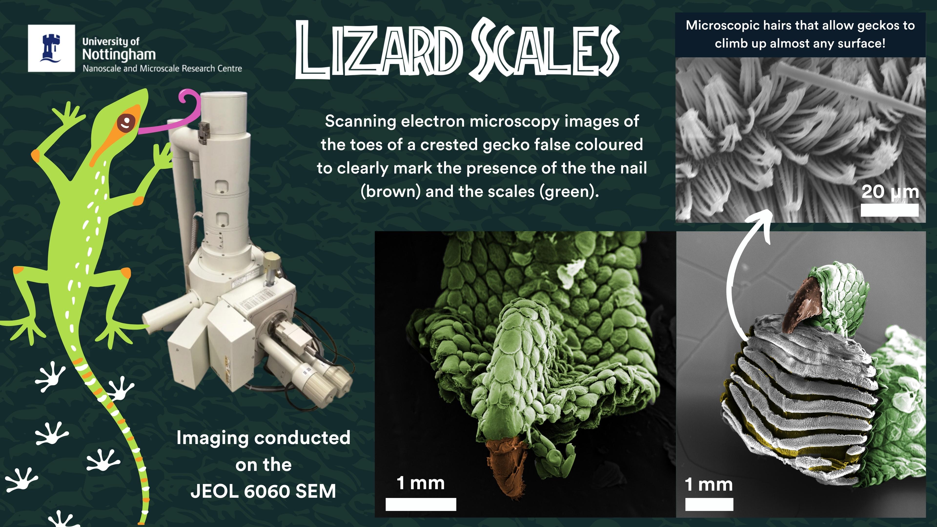
Lizard Scales - August 2023
Delve into the microscopic realm of a crested gecko's toe with our latest Under the Microscope suggestion...Lizard Scales! SEM imaging showed the presence of the scales as well as the microscopic hairs that allow geckos to climb up almost any surface. Imaging was conducted on a JEOL 6060 SEM by Tom Hartman (School of Life Sciences) and Alice Campain. Thanks to Tabitha for the suggestion!
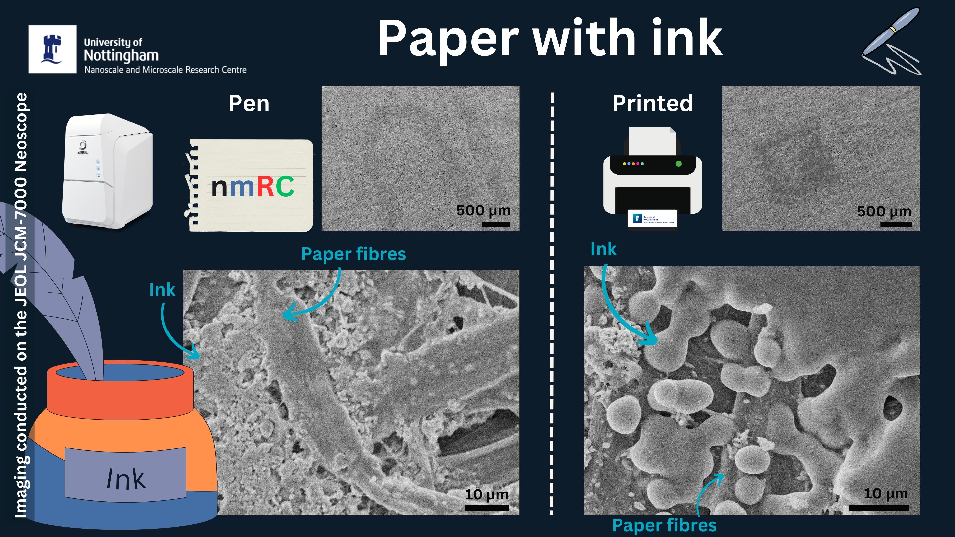
Inked paper - July 2023
Have you ever wondered what inked paper looks like at the microscale? Our latest Under the Microscope winner Andrei had that very same thought and decided to submit the idea to us, and we were excited to image it! We produced two inked paper samples; 1) written letters ("nmRC") using a ballpoint pen, and 2) a printed University of Nottingham castle logo. Both samples were then imaged using Scanning Electron Microscopy to observe any differences. In both high magnification images the paper fibres can be observed, but the ink produced by the printer appears far more globular than that of the pen which appears more grainy, and sand-like! Imaging was conducted using a JEOL JCM-7000 Neoscope SEM operated by Dr Luke Norman (Knowledge Exchange Fellow).
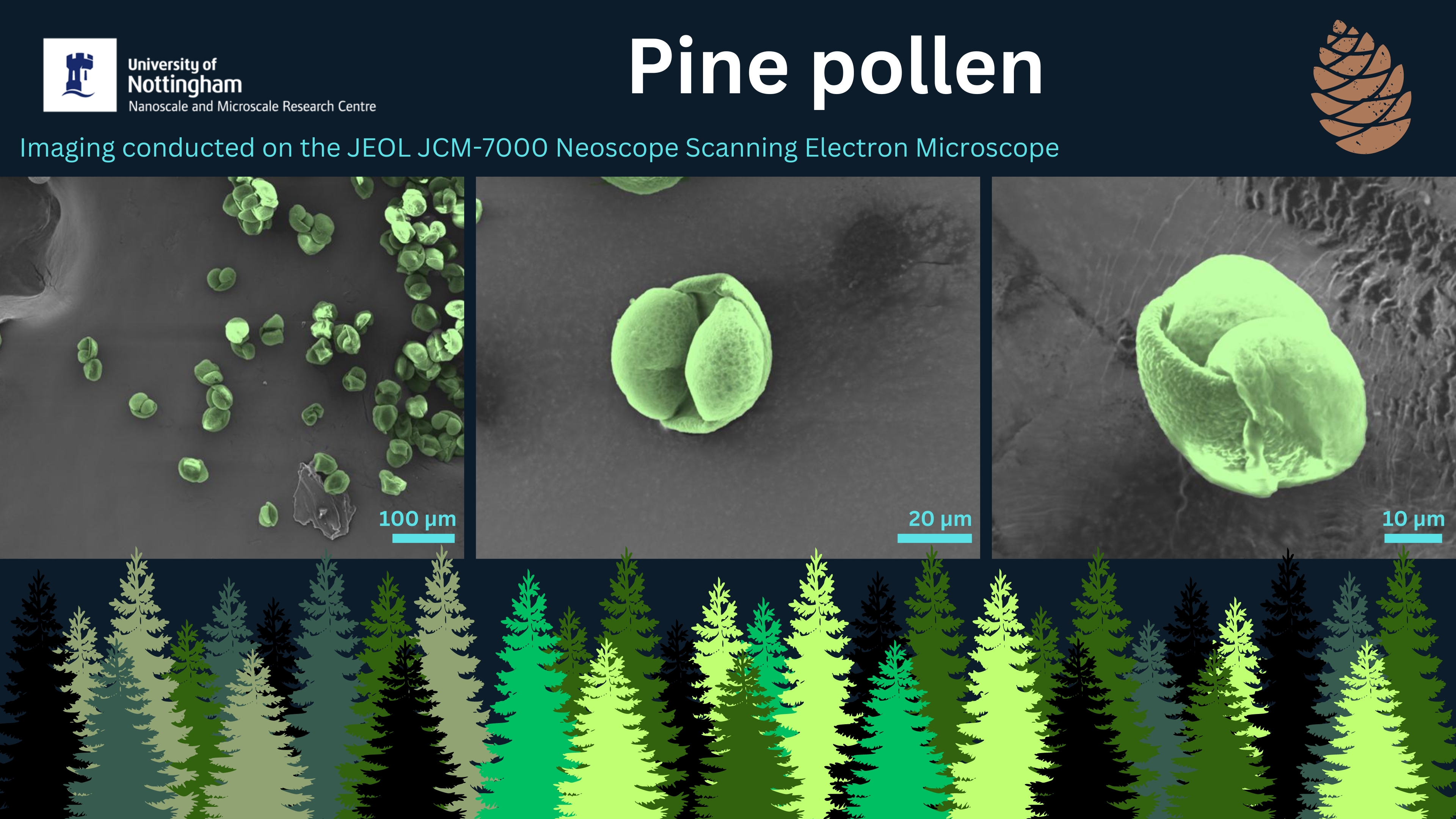
Pine Pollen - June 2023
It's hayfever season and there is lots of pollen about, but have you ever wondered what pollen looks like at the microscale? Clare Templey suggested for us to image pine pollen using electron microscopy and we thought it was a great idea! The pollen was collected from some pine trees around University Park campus and then analysed using the JEOL JCM-7000 Neoscope Scanning Electron Microscope by Dr Luke Norman. If you're inspired by this suggestion and would like to nominate something for us to image, let us know!
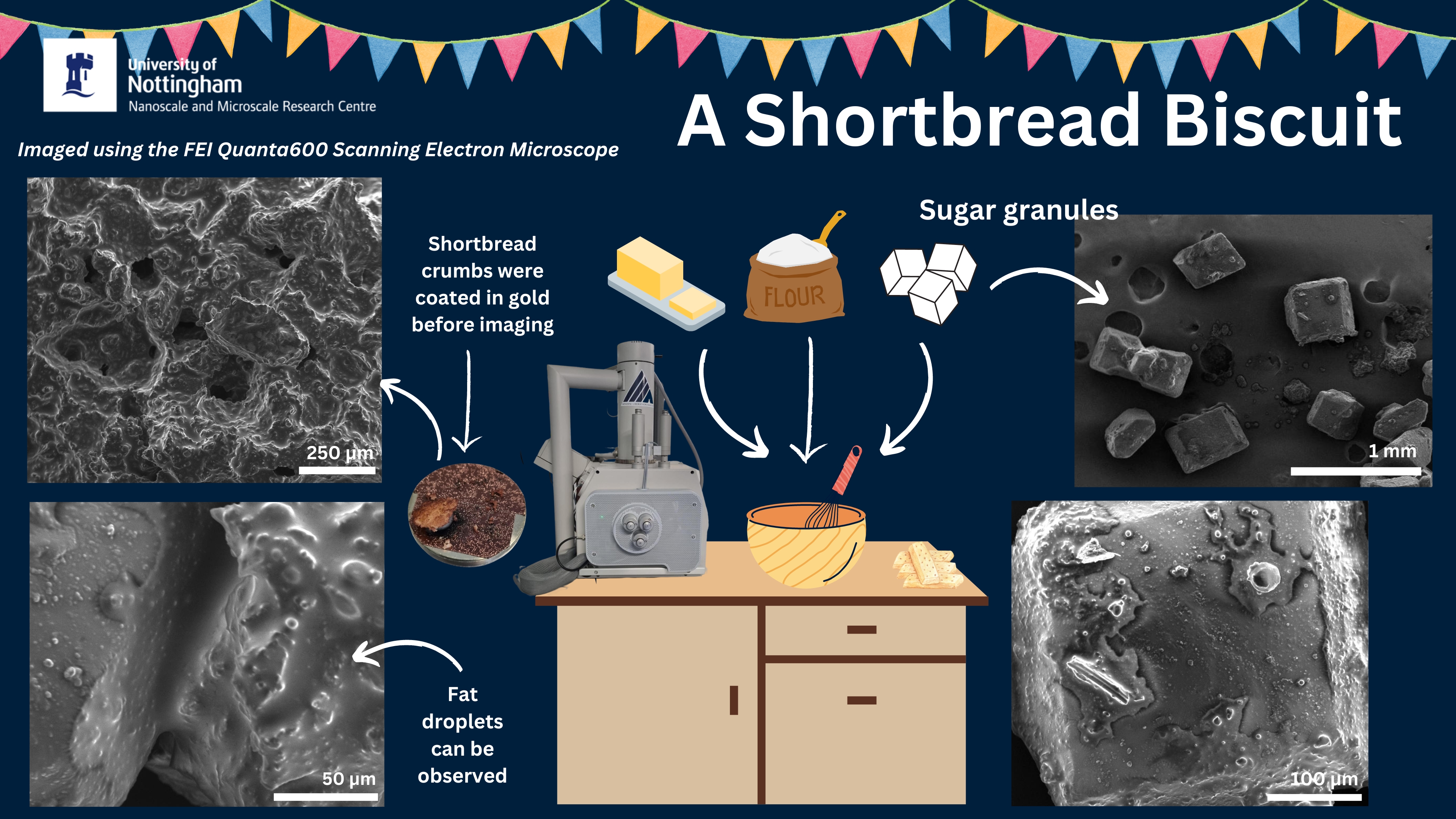
A Shortbread Biscuit - May 2023
Calling all baking enthusiasts and curious foodies! Our May 2023 Under the Microscope winning entry is... A Shortbread Biscuit submitted by Taranvir Bedi. Imaging was conducted using a Thermo Fisher (FEI) Quanta600 Scanning Electron Microscope by Lorelei Robertson. Various shortbread crumbs were analysed as well as some sugar granules that were on top. Prepare to be amazed as we unveil the intricate microstructure of everyone's favourite baked treats,and don't forget to suggest your ideas to be in with a chance to be chosen next month!
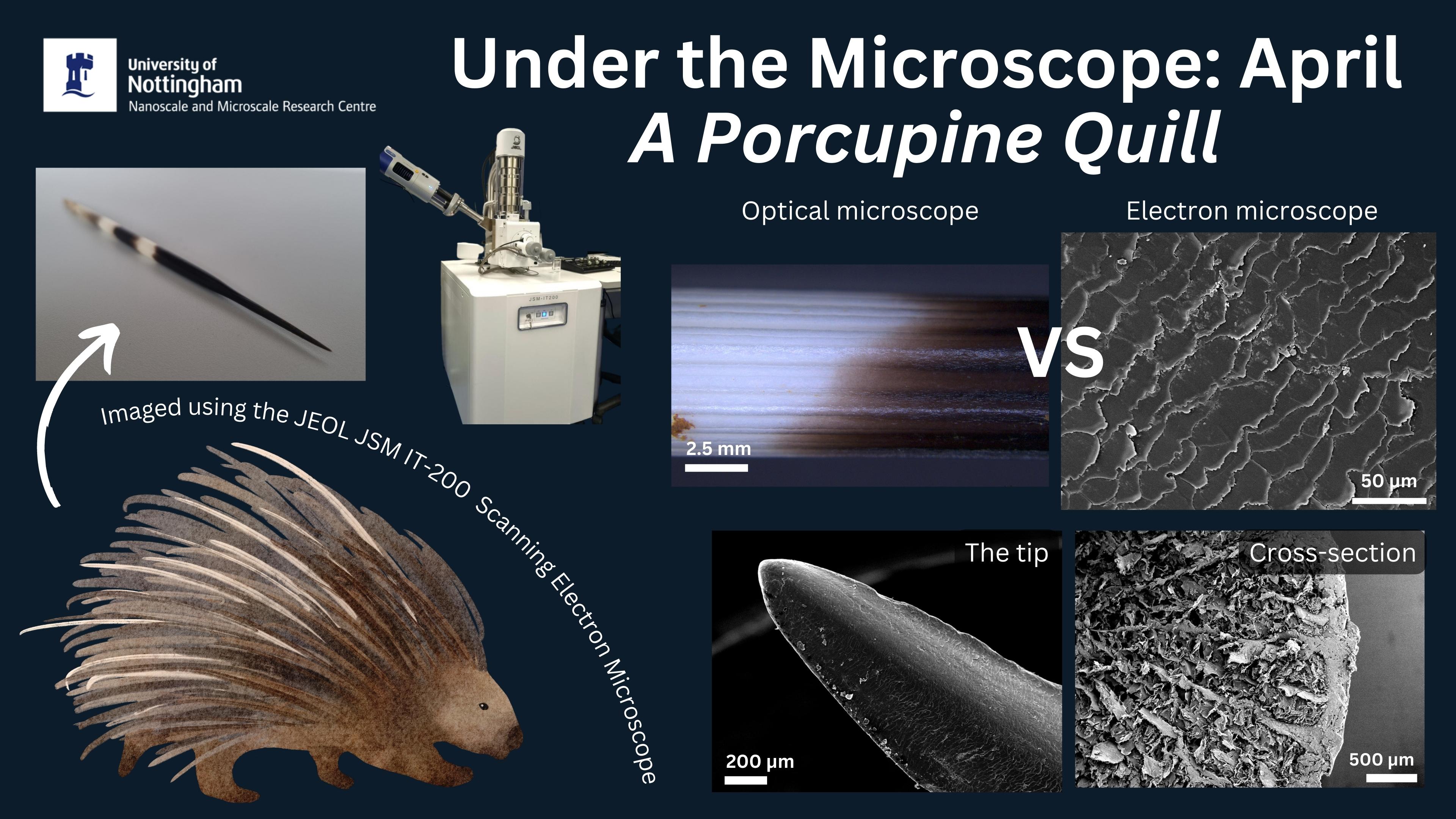
A Porcupine Qull - April 2023
The winning entry for April 2023 was a porcupine quill which was suggested by Susannah Goh who picked this idea because "Porcupines are always rather fun and interesting in all of their aspects". Imaging of the tip, exterior and cross-section was conducted by Lorelei Robertson on the JEOL IT-200 SEM. Check out the images below, and don't forget to suggest your ideas to be in with a chance to be chosen next month!
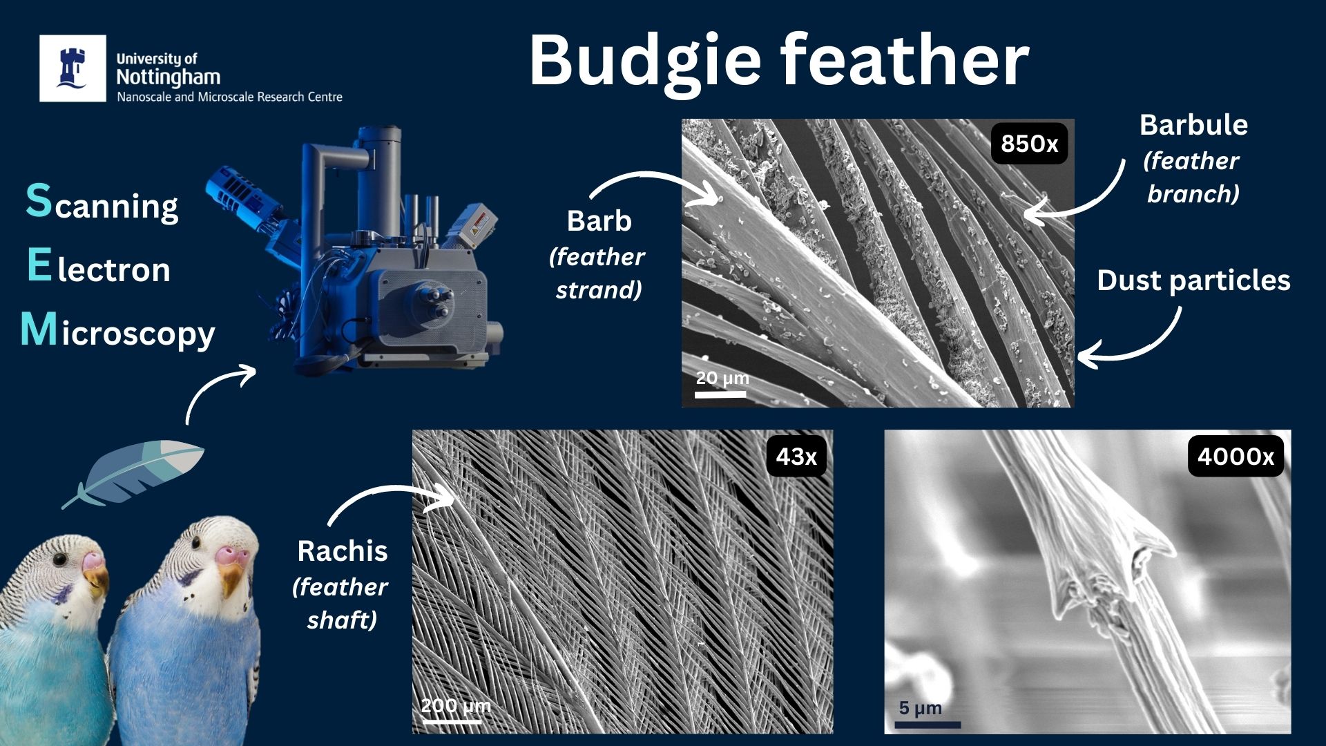
Budgie Feather - March 2023
During a visit by NottsTV, their producer bought in a feather from her pet budgie called Dudley to image by SEM. The feather was first coated in gold to make it conductive and then was imaged by Lorelei Robertson on the JEOL IT-200 SEM. Results below...
Example Materials Imaged
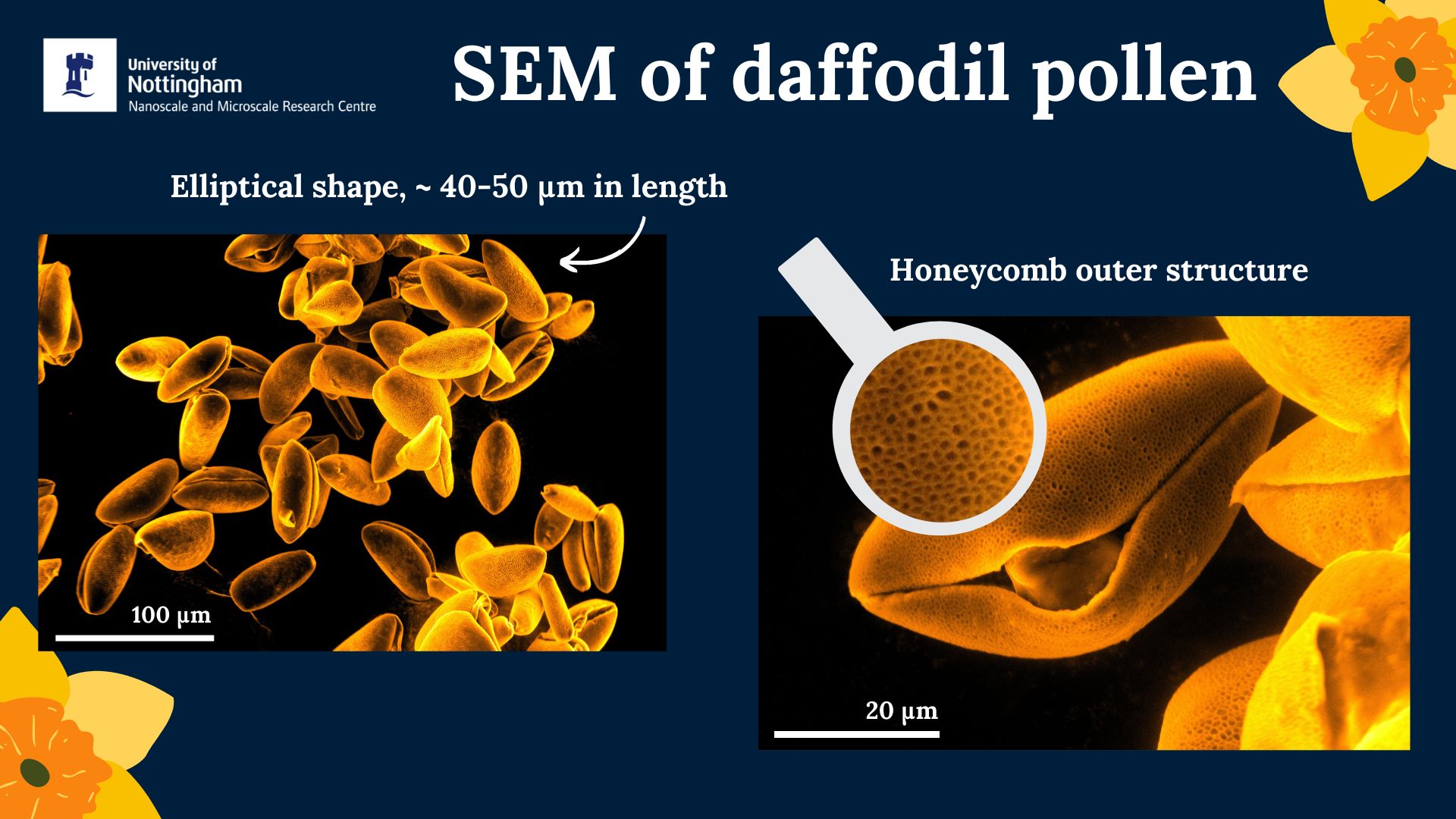
Daffodil Pollen
Daffodil pollen was chosen to celebrate St David's Day in Wales, and was imaged by Dr Beth Steer. The images revealed a honeycomb outer structure to the pollen!
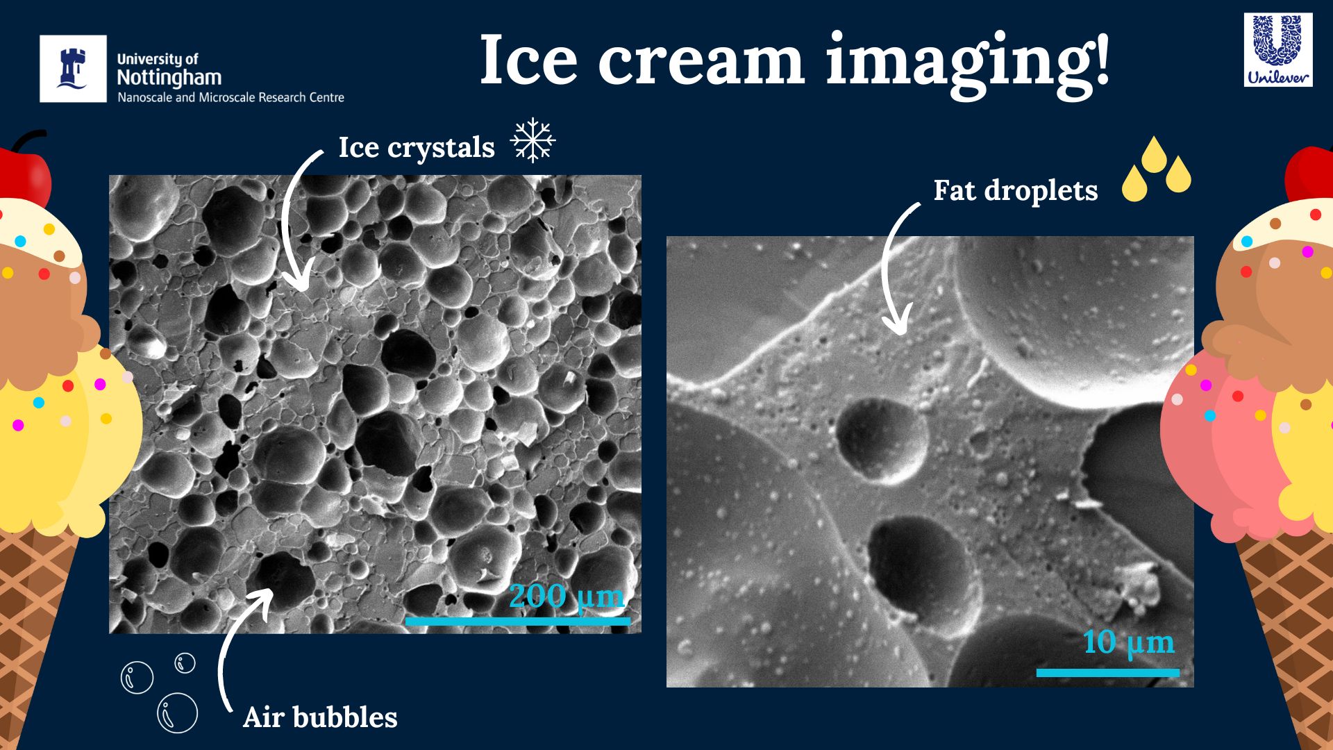
Ice Cream
The microstructure of ice cream was imaged by Dr Chris Parmenter, and identified three main components which were: ice crystals, air bubbles, and fat droplets.