The laboratory is equipped with a 275 channel CTF whole head MEG system capable of measuring neuromagnetic fields, genetared by the brain, on the fT scale at sampling rates up to 19,000Hz.
Coregistration of the MEG sensor locations to MRI defined brain anatomy is achieved using a 3D head digitisation. This ultimately facilitates 3D images showing moment to moment changes in brain current density.
CTF 275 channel MEG Scanner
Dr Matt Brookes using the CTF 275 channel MEG scanner
Capabilities
MEG compatible EEG
Concurrent MEG and EEG recordings are made possible by integration of a brain products 64 channel MEG compatible EEG system into the MEG system.
This allows recording with an effective 339 channel MEG/EEG system. In addition to EEG, electrode channels coupled to the MEG also enable simultaneous recording of ECG, EMG and EOG.
Stimulus Presentation
The lab is equipped to deliver most forms of stimulation including visual, auditory and somatosensory (including median nerve and vibrotactile stimulation).
A separate stimulus computer is used to present paradigms to the subject and this is equipped with MATLAB (Cogent toolbox), PsychoPy and Presentation (Neurobehavioural systems) for paradigm generation.
Data Processing
Data processing can be carried out using a number of high end PC’s with a Linux operating system, available for use at the Sir Peter Mansfield Imaging Centre.
Software packages available to process MEG data include CTF commercial software (incorporating Synthetic Aperture Magnetometry (SAM)), SPM8, fieldtrip and NutMEG.
In house Matlab programs are available comprising custom written beamformer and minimum norm source localisation algorithms.
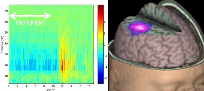
Changes in electrical brain activity induced by finger movement. The right hand image shows the spatial signature of beta band oscillatory modulation. The left hand image shows the time frequency representation.
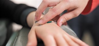
Electromyography electrodes being attached to a person to allow the relationship between muscle activity and brain activity to be observed
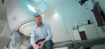
Assistant Professor Dr Matt Brookes in front of the CTF 275 channel MEG scanner