High Content Screening
High content screening is a range of methods used to evaluate multiple biochemical and morphological parameters in cellular systems. A wide range of cells cultured inside microtitre plates (6 to 1536 wells) can be imaged. Combining quick automated and reliable image acquisition with powerful image analysis algorithms it is possible to automate and quantitative many biochemical and morphological properties at the single cell level. These methods can be used in a wide range of research, but shows the most strength in drug discovery where it is becoming an indispensable tool to compound screening. The facility is part of the drug discovery platform.
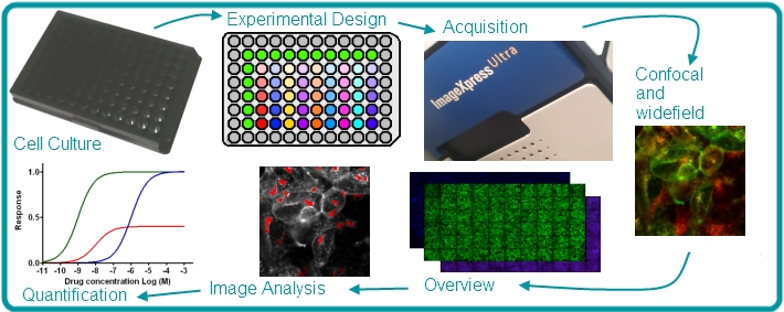
The ICS high content screening service boasts two plate imaging platforms, ImageXpress Ultra, a true point scanning confocal microscope and the ImageXpress Micro, a fluorescence widefield system with environmental control and drug robot. The service also has analysis algorithms from Molecular devices (MetaXpress) and a dedicated image analyst.
For more information, please contact Tim Self: 0115 823 0090 or Josephine Gilbert: 0115 823 0080
Molecular Devices ImageXpress Micro
 The micro is a environmentally controlled widefield/epifluorescent system with fluidics allowing for extremely flexible live cell work, as well as fixed cell imaging. The fluidics allow for drug addition without removing the cells from the microscope. It is optimised for speed, with a laser autofocus system and a Xenon lamp illumination. It is compatible with slides and plates from 6 to 1536 wells.
The Micro has four objective lenses:
| Magnification | Type | Numerical Aperture |
|---|
| 10x |
Plan Fluor |
0.30 |
| 20x |
Plan Fluor Extra Long Working Distance |
0.45 |
| 40x |
Plan Fluor Extra Long Working Distance |
0.60 |
| 60x |
Plan Fluor Extra Long Working Distance |
0.85 |
For more information, please contact Tim Self: 0115 823 0090 or Josephine Gilbert: 0115 823 0080
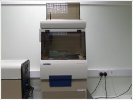
Molecular Devices ImageXpress Ultra
The Ultra is a fully integrated true point scanning confocal microscope for automatic acquisition for high content cell based screening. The system houses four solid state lasers, providing 4 channel excitation of flourophores at the most frequently used wavelengths and four detector photomultiplier tubes (PMT) for the collection of images. Those PMT’s have four bandpass filters, covering most common use dyes.
The available PMT and Laser lines are:
| PMT | Excitation Laser (nm) | Emission Bandpass Filter (nm) | Common Dyes |
|---|
| 1 |
405 |
477/60 |
DAPI |
| 2 |
488 |
525/50 |
FITC |
| 3 |
561 |
593/40 |
Texas Red |
| 4 |
635 |
685/40 |
Cy5 |
The ultra has 5 air objective lenses:
| Magnification | Type | Numerical Aperture |
|---|
| 10x |
Plan Fluor |
0.30 |
| 20x |
Plan Fluor Extra Long Working Distance |
0.45 |
| 40x |
Plan Apo |
0.95 |
| 40x |
Plan Fluor Extra Long Working Distance |
0.60 |
| 60x |
Plan Fluor |
0.85 |
The ultra can scan all standard microtitre plates and slides. The ultra can also be used with the KiNEDx selective compliant assembly robot arm (SCARA) to fully automate multiple plate acquisition on microtitre plates.
For more information, please contact Tim Self: 0115 823 0090 or Josephine Gilbert: 0115 823 0080
KiNEDx Plate Handling Robot
The KiNEDx selective compliant assembly robot arm (SCARA) is a plate handling robot that can store, load and remove plates from the hotel to/from the ImageXpress Ultra. The SCARA can handle up to 48 plates (with or without lids). This allows for fully automated high throughput screening with the ultra and removes the need for manual handling of plates.
For more information, please contact Tim Self: 0115 823 0090 or Josephine Gilbert: 0115 823 0080
Molecular Devices Flexstation 1 and 3
We have two Molecular Devices Flexstations 1 and 3, both are scanning fluorometers with integrated 8 channel pipettors for fluid transfer, useful for measuring end point or changes in absorbance or intensity (when using fluorescence). The flexstation can measure fluorescent intensity and changes in florescence, which can be used in assays such as calcium flux and membrane potential, as well as many others. The Flexstation 3 has enhanced capability for luminescence reads. With a temperature control of 2oC above ambient to 45oC it provides a very flexible live cell platform.
For more information, please contact Tim Self: 0115 823 0090 or Josephine Gilbert: 0115 823 0080
Image Analysis
We have 7 algorithms for use in the MetaXpress software that automatically analyses the data taken from the ImageXpress Ultra and ImageXpress Micro:
Multi-wavelength cell scoring
Used for counting cells and logging measurements in two or three channel experiments.
Granularity/Transflour
Used for experiments such as analysis of receptor internalisation and the quantitation of intensity of structures e.g. nuclei, pits or vesicles.
Nuclear Translocation HT / Translocation / Translocation and Translocation-Enhanced
Used for quantitation of correlation between probes or compartments. Measurements include mean compartment area, correlation coefficient, intensity and number / percentage of positive cells.
Cell cycle
Can be used for the quantitation of cell cycle stages with apoptosis and mitosis specific measurements.
There are other image analysis suites routinely used within the high content screening facility. Please contact us regarding your specific requirements.
For more information, please contact Tim Self: 0115 823 0090 or Seema Rajani: 0115 823 0080