Raman Spectroscopy
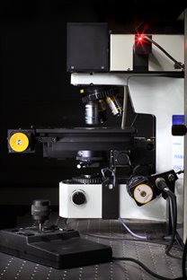
The HORIBA LabRAM HR confocal Raman microscope available at the nmRC
Raman Spectroscopy at a glance
Raman spectroscopy is a non-invasive optical technique used to determine the chemical identity and state of a sample by its unique vibrational (molecular) modes. Inelastic (loss or gain of energy) scattering of light from the sample is measured, with distinct shifts in the energy of the scattered photons being representative of the sample chemistry.
Applications
- Vibrational spectra of substrate chemistry
- Confocal component mapping and depth profiling
- UV-Visible-IR excitation analysis
- Temperature and time dependent chemical and physical state change identification
- Ambient or controlled environment analysis
How does Raman work?
Raman spectroscopy and microscopy is a uniquely sensitive analytical technique that is non-invasive in nature. It can rapidly provide quantifiable identification of bulk and surface species with exceptional resolution. The technique measures the interaction between a monochromatic light source and the vibrational (and other low-frequency) modes of the molecular systems being analysed.
A given molecular vibration can be excited by a certain frequency of incident light to a higher virtual energy state. This higher energy state will almost always relax again to a lower energy state. While normally this occurs elastically (to its original energy state as Rayleigh scattering) it may also occur inelastically. This represents relaxation to a different vibrational energy level, generating the release of a photon with an altered frequency to that of the incident photon.
This change in frequency is the Raman shift, and can be recorded and plotted against the scattered light intensity. Therefore Raman scattering gives rise to reductions or gains in light frequencies that correspond very sensitively to the molecular structure and state of the material being analysed. Solving these spectra allows characterisation of the chemical and physical state of a material.
Our Raman Spectroscopy Facilities
- Four excitation wavelengths available:
- Kimmon IK3201R-F He-Cd laser (λ = 325 nm, average power = 20 mW)
- Laser Quantum Torus 532 100mW laser (λ = 532 nm, average power = 129 mW)
- Laser Quantum Ignis 660 100mW (λ = 660 nm, average power = 187 mW)
- Toptica Photonics XTRA-PS (λ = 785 nm, average power = 385 mW)
- Choice of standard CCD (HORIBA Synapse) or EM-CCD (Andor Technology Newton 970).
- Olympus BX41 stage with x40 (UV only), x10 and x50 (visible and NIR) objective lenses.
- Linkam Scientific TMS 94 system (thermal stage) capable of operating between -196 and 350 ˚C.
- SWIFT® mode offers confocal Raman mapping with CCD integration times down to 1 ms per point and below.
- DuoScan® mode is a confocal imaging mode, with high precision, ultra-fast rastering mirrors creating variable sized laser macro-spots, and also allowing nano-step mapping from deep UV to NIR.
- Choice of gratings for standard or high resolution spectroscopies.
HORIBA XploRA INV Inverted Raman Microscope
- Inverted microscope geometry suitable for 'in-situ' analysis and biological samples.
- Excitation wavelength available: 785nm.
- Gratings available: 600, 1200, 1800 and 2400 lines/mm (configuration dependent).
- Objectives available: 10x, 20x and 100x.
HORIBA LabRam HR Evo Nano (DCI-TERS)
Scanning probe microscope coupled to a Raman microscope enabling tip-enhanced Raman spectroscopy (TERS) and co-localised AFM-Raman.
- Conventional upright microscope geometry.
- Excitation wavelengths available: 532, 633 and 785 nm.
- Gratings available: 150, 600, 1800 and 2400 lines/mm.
- Objectives available: 5x, 10x, 50x, and 100x.
- Controllable confocality for standard or high spatial resolution imaging.
- Motorised half-wave and analyser plates for polarised Raman measurements.
- Ultra-low frequency (ULF) Raman module allowing measurement in the sub-50 cm-¹ region.
- Imaging modes include AFM (contact, semi-contact, non-contact), KPFM, SCM, EFM, PFM, cAFM, LFM, FMM, MFM, STM and nanolithography.
- Dual optical access from the side and below for reflectance and transmission TERS measurements.
- Protective enclosure for environmental control.
- Sample and cantilever holders for variable temperature analysis in air (-50 to +300 oC) and liquids (ambient to +60 oC).
- Electrochemical cell for ecTERS.
Publications of Interest
- A one-pot-one-reactant synthesis of platinum compounds at the nanoscale, T., Biskupek, J., Li, Z. Y., Rance, G. A., Botos, A., Fogarty, R. M., Bourne, R. A., Yuan, J., Lovelock, K. R. J., Thompson, P., Fay, M. W., Kaiser, U., Chamberlain, T. W. & Khlobystov, A. N., Nanoscale, 9, 14385–14394 (2017).
- Growth of Carbon Nanotubes inside Boron Nitride Nanotubes by Coalescence of Fullerenes: Toward the World’s Smallest Coaxial Cable, Walker, K. E., Rance, G. A., Pekker, Á., Tóháti, H. M., Fay, M. W., Lodge, R. W., Stoppiello, C. T., Kamarás, K. & Khlobystov, A. N., Small Methods, 1, 1700184 (2017).
- Host–Guest Hybrid Redox Materials Self-Assembled from Polyoxometalates and Single-Walled Carbon Nanotubes, Jordan, J. W., Lowe, G. A., McSweeney, R. L., Stoppiello, C. T., Lodge, R. W., Skowron, S. T., Biskupek, J., Rance, G. A., Kaiser, U., Walsh, D. A., Newton, G. N. & Khlobystov, A. N., Advanced Materials, 31, 1904182 (2019).
- Direct Synthesis of Multiplexed Metal-Nanowire-Based Devices by Using Carbon Nanotubes as Vector Templates, Clément, P., Xu, X., Stoppiello, C. T., Rance, G. A., Attanzio, A., O’Shea, J. N., Temperton, R. H., Khlobystov, A. N., Lovelock, K. R. J., Seymour, J. M., Fogarty, R. M., Baker, A., Bourne, R. A., Hall, B., Chamberlain, T. W. & Palma, M., Angewandte Chemie International Edition, 58, 9928–9932 (2019).
- Gel–Polymer Electrolytes Based on Poly(Ionic Liquid)/Ionic Liquid Networks, Sen, S., Goodwin, S. E., Barbará, P. V., Rance, G. A., Wales, D., Cameron, J. M., Sans, V., Mamlouk, M., Scott, K. & Walsh, D. A., ACS Applied Polymer Materials, 3, 200–208 (2021).