Our Facilities and Expertise
The nmCS team offer access to a huge variety of surface analytical facilities both within the nmRC itself and also across the wider University. We have a wealth of world leading instrumentation and expertise in materials characterisation, specialising in imaging and compositional analysis.
We also have a selection of e-leaflets for most of our equipment which highlight key capabilities, instrumentation, and relevant case studies, as well as a facilities brochure which can be found here: nmRC Facilities Brochure.
For more information regarding our core facilities and to explore how we can help you better access and perform them please see below. Click on the header to expand each section and learn a little bit more about the theory behind the techniques, the instrumentation we possess and what it could do for you. At the bottom of the page you will find a series of e-leaflets containing capabilities of most of our facilities. For any further information or queries please get in touch.
e-leaflet available here
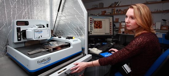
Atomic force microscopy (AFM) is an example of high resolution scanning probe microscopy, which allows the imaging and physicochemical analysis of molecular surfaces with nanometre resolution.
e-leaflet available here
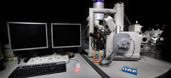
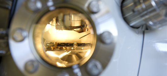
SEM is an imaging technique with high depth of field and lateral resolution. It uses electrons to generate secondary sample irradiance. This can then be analysed to visualise sample surfaces as well as analyse the physical and chemical state of the substrate.
e-leaflet available here
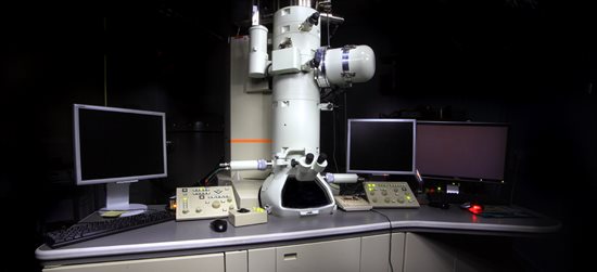
Transmission Electron Microscopy (TEM) is capable of providing very high resolution images down to a level of several Angstroms (~ 0.19nm). The study of nano-scale morphological and chemical features in cells or different materials down to near atomic levels is possible.
e-leaflet available here
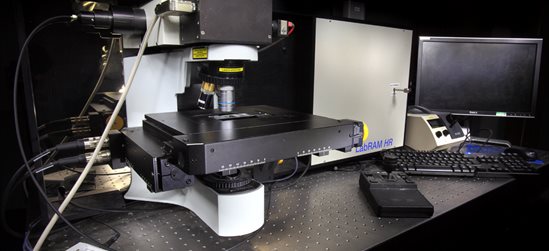
Raman spectroscopy is a non-invasive technique that generates a specific fingerprint spectrum through which the molecule or compound can be identified or observed in real-time transitions.
e-leaflet available here
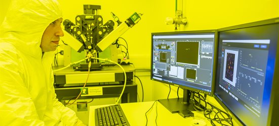
Secondary Ion Mass Spectrometry (SIMS) is a highly sensitive surface analytical method that can describe the chemical character of a substrate surface in 3D. Solid surfaces can be analysed by either a high mass resolution Orbitrap analyser or high imaging resolution time of flight (ToF) analysers.
e-leaflet available here
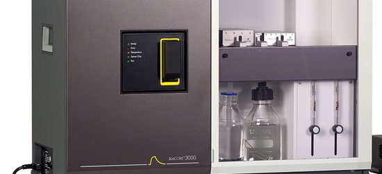
Surface Plasmon Resonance (SPR) allows sensitive detection of molecular interactions in real time without labels. It can measure the binding, kinetics, affinity, specificity and concentration of an event and saves the work of purifying and labelling materials.
e-leaflet available here
Ellipsometry is an optical technique used to determine thin film thicknesses with Ångström resolution and infer material properties.
e-leaflet available here
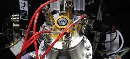
X-ray photoelectron spectroscopy (XPS) is a surface analysis technique that qualifies and quantifies the elemental composition and chemical state of a material.
e-leaflet available here
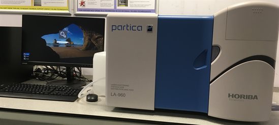
Particle size analysis is the characterisation of the size distribution (size range and/or mean size) of particles in a sample. Particle sizing can be applied to solid materials, suspensions, emulsions and aerosols.
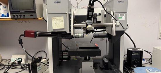
Measuring the contact angle of a liquid on a solid surface allows quantification of the wettability (how a liquid spreads). This can in turn be used to investigate the energetics of an interface.
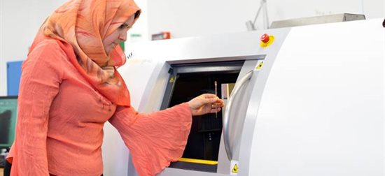
X-ray Computed Tomography (CT) is a non-invasive, non-destructive imaging technique permitting the visualisation and quantification of the interior structure of an object in three dimensions. Micro-CT generates cross-sections with pixel sizes in the micrometre range for high resolution imaging.
e-leaflet available here
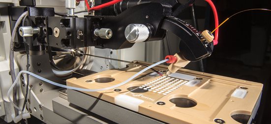
LC-MS is a versatile and highly sensitive analytical technique for the measurement of small molecular weight compounds in a diverse range of sample types. It uses a series of mass detection systems to provide both quantitative and qualitative analyses.
Fluorescent nanosensors are spherical probes composed of an inert matrix with nanometre sized dimensions that selectively respond to stimuli in their surroundings to transduce fluorescence signals to a detector.
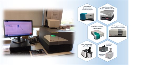
Spectroscopy involves an interaction between light (electromagnetic radiation) and matter, including absorption, emission, scattering, refraction, resonance and diffraction.
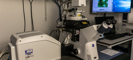
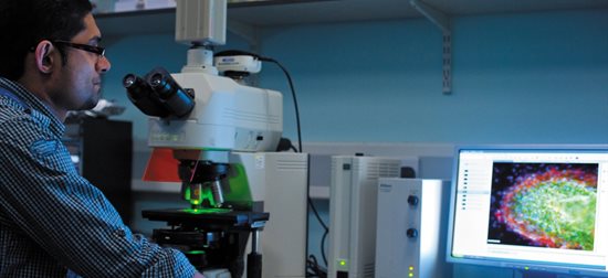
Confocal Laser Scanning Microscopy (CLSM) is a light microscopy technique used to capture high resolution images of samples including cells and other biological species.
nmRC Facilities Brochure
Our facilities brochure highlights the key techniques that nmCS offers, and outlines the capabilities and applications of each one as well as some relevant case studies.
To get your copy: either click the link below or pop into the centre for a physical copy. They can be found in our entrance foyer.
nmRC Facilities Brochure