Quantitative MRI to assess portal hypertension in cirrhosis patients at 3T
Hepatic venous pressure gradient (HVPG) is the gold standard method for the assessment of portal pressure, but highly invasive. We scanned patients with portal hypertension at both 1.5T and 3T to assess MRI parameters related to portal pressure as defined by HVPG. Iron-corrected liver T
1 highly correlated over the full range of HVPG (3T p<0.0002, 1.5T p<0.0001), spleen T
1 and superior mesenteric artery velocity correlated up to HVPG of 15 mmHg (spleen T
1: 3T p<0.0003, 1.5T p<0.0006; SMA velocity: p<<0.00001), after which at HVPG >15 mmHg no correlation was observed.
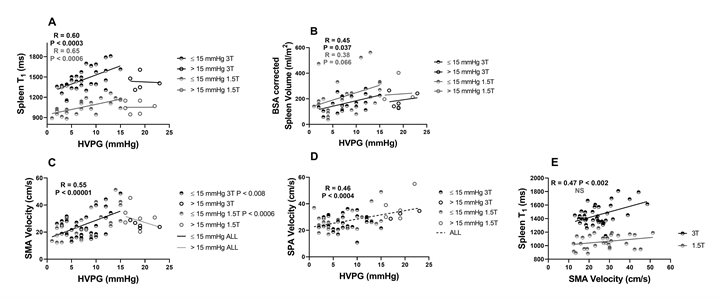
Figure: A Spleen T1 correlated with hepatic venous pressure gradient (HVPG) up to 15 mmHg at 1.5T (R=0.65, P<0.0006) and 3T (R=0.60, P<0.0003); B Body surface area (BSA) corrected spleen volume correlates with HVPG up to 15 mmHg for 3T (R=0.45 P=0.037) and 1.5T (R=0.38, P<0.066) C Superior mesenteric artery (SMA) velocity correlates with HVPG up to 15 mmHg for 1.5T and 3T data combined (R=0.55, P<0.00001), after which SMA velocity reduces; D Splenic artery (SPA) velocity correlated with HVPG (R=0.47, P<0.002); E Correlation of spleen T1 with SMA velocity at 3T (R=0.47, P<0.002).