Infastructure
Expertise and Equipment in the Division of Advanced Materials and Healthcare Technologies
Infastructure
Atomic Force Microscopy
We have a wide range of state-of-the-art Atomic Force Microscopes in the laboratory, providing high-quality surface imaging and analysis for various biomolecular and pharmaceutical applications. The following are a few highlights.
Combined ICON/FastScan_Bio Atomic Force Microscope
Bruker
This latest AFM features three new technologies from Bruker, ScanAsyst™, PeakForce™ and QNM™. The PeakForce™ tapping mode provides a direct force control with minimum peak force down to less than 100pN. The QNM™ (Quantitative Nanomechanical Measurement) mode provides simultaneous high-resolution and well-defined force mapping (localized and quantitative elastic modulus, adhesion, deformation, dissipation, etc.) With the ScanAsyst™, the user can easily operate the AFM in a completely “automatic” mode for the first time. Whilst ICON can provide 100µm x 100µm x 10µm large X-Y-Z scope imaging, FastScan_Bio can provide 1-second per frame fast imaging in air or liquid. In addition to this ICON/FastScan_Bio AFM, we have an army of various AFM instruments, including MultiMode, Dimension, eScope and Scanning Thermal AFMs from Bruker, MFP-3D from Asylum Research and ForceRobot from JPK.
nano-TA2™
Anasys Instruments
The new generation of thermal probes can image the sample of interest with sub-30nm spatial resolution (in contact or intermittent contact modes) and identify the regions to perform Localized Thermal Analysis, similar to DSC (Differential Scanning Calorimetry) measurement but localized to a surface area less than 100nm in diameter.
ForceRobot® 300
JPK Instruments
This latest fully automated molecular force spectroscope also provides the highest flexibility. Users can design complex experimental details (fluids, temperatures, force ramp parameters, etc.) of unlimited number of force measurement in advance and leave the instrument to complete the job automatically.
EnviroScope Atomic Force Microscope
Bruker
This special AFM allows observation of sample properties under precise temperature and humidity control. In addition to this Dimension-based eScope AFM, we also have heating stages for our MultiMode AFM instruments.
Top
Cantilever Sensor
Cantisens® Research installed in LBSA is designed to support leading edge scientific research and industrial application development giving users access to innovative Cantilever Sensor technology.
Using customized chemical sensing layers, Cantisens® Research can be employed for a wide range of scientific and industrial applications.
The stand-alone system offers parallel, label-free detection of up to eight different substances or biomolecular interactions. It combines a multi-purpose cantilever sensor platform with powerful measurement electronics and easy-to-use control and evaluation software.
More information about the instrument and the Cantilever Sensor technology can be found on the Concentris website.
Top
Confocal Microscopy
Leica TCS SP confocal microscope is installed in LBSA. In contrast with the conventional wide-field fluorescence microscope, where the entire specimen is illuminated by a light source, a confocal microscope uses point illumination and a pinhole in an optically conjugate plane in front of the detector to eliminate out-of-focus information. Only the light within the focal plane can be detected, resulting in much better image quality than that of wide-field images.
Please see the Leica website for more information on confocal microscopy.
Top
Contact Angle Measurement
Both micro and pico litre liquid contact angle measurements are routinely made within the LBSA. A KSV instrument is used to make the former measurements with manual dispensing and automated image analysis of the drop. The automated pico litre dispenser is used to make measurements on a length scale around one tenth of 1 mm and is fully automated.
See, for example, Taylor, M.; Urquhart, A. J.; Zelzer, M.; Davies, M. C.; Alexander, M. R., Pico litre water contact angle measurement on polymers. Langmuir Letters 2007, 23, (13), 6875-6878
Please see the KRÜSS website for technical details.
Top
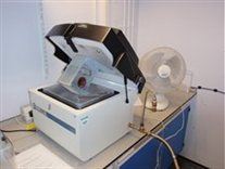
Differential Scanning Calorimeter
The Q2000 Differential Scanning Calorimeter (DSC) was delivered in mid 2009. This state of the art DSC has an automated sample change robot and a 50 sample magazine. It is capable of heating and cooling rates from 0.1 to 200 C per minute, and can run in both standard and temperature modulated modes, as well as quasi-isothermal.
It stores up to 10 calibrations and is ideal for high-throughput, high precision measurements of the thermal properties of materials from -40 to 600 C. Typically, around 1 mg of sample is required.
Top
PriorLux Polarised Light Microscope
The PriorLux polarised light microscope operates in transmission and is equipped with a Linkam DSC600 hot-stage. It was installed in late 2009. It allows simultaneous recording of a polarised optical video and a DSC trace. It is routinely capable of a temperature range -196 to 600 C. It requires only microscopic amounts of sample for optical measurements, and around 1 mg for concomitant DSC characterisation. It is used as a powerful complementary tool to the Q2000 DSC.
Top
Quartz Crystal Microbalance
The QCM-D (Quartz Crystal Microbalance with Dissipation monitoring) is based on a technology developed by Q-Sense by collecting both the dissipation and the resonance frequency of a quartz crystal. It can be used to study the formation of thin films such as proteins, polymers and cells onto surfaces in liquid.
Technical details of QCM-D can be found on the
Q-Sense website.
Top
Scanning Thermal Microscopy
If the normal solid sharp probe used in AFM is replaced with a ultra miniaturised resistance, the image contrast reflects variations in thermal properties across the sample surface. The probe can be used as either a micro-thermometer to obtain a temperature "map", or a highly localised heater to obtain a conductivity/diffusivity "map" of the sample surface.
This equipment can also be used to measure localized thermal transitions, similar to the measurement done by the traditional DSC but in a very small surface domain.
In addition to the old generation of SThM with a micro-thermal probe attached to one of our Explorer AFMs, the newnano-TA2 from Anasys Instruments has also installed on one of our MultiMode AFMs. The proprietary thermal probes used in nano-TA2 can achieve scanning thermal microscopy and local thermal analysis with sub-100 nm resolution.
Top
Spex6870 Cryomill
The Spex 6870 cryomill is used to mill materials at liquid nitrogen temperature, and was commissioned in late 2009. It is used to produce a wide range of amorphous materials, up to 100 g. Milling renders materials amorphous, and the low temperatures prevent thermally-driven recrystallisation.
We also have a Retsch MM400 mixer ball mill (not shown) which operates at room temperature and is complementary to the cryomill.
c
Top
Surface Plasmon Resonance
Surface Plasmon Resonance (SPR) allows sensitive detection of molecular interactions in real time without the use of labels. This saves the work of purifying and labelling material, and eliminates the risk that labels may interfere with the interaction being studied. Biacore 3000 is the highest performance research system available for label-free studies of biomolecular binding.
Please see
Biacore's webpage for specification details of Biacore 3000.
Top
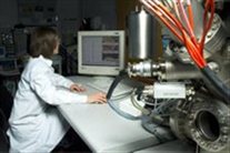
Time of Flight Secondary Ion Mass Spectroscopy and X-ray Photoelectron Spectroscopy
The state-of-the-art Time of Flight Secondary Ion Mass Spectrometer (ToF-SIMS) IV (ION-TOF GmbH, Münster, Germany) is equipped with a liquid metal (Bi) ion gun (LMIG) for spectroscopy and chemical imaging. In addition, an electron impact ion source allows spectroscopy using polyatomic primary ions and depth profiling using either C60+ or Cs+ (combined with crater analysis using the LMIG or C60+). The reflectron ToF mass an alyser has a mass resolution in excess of 10,000.
Upgrades
Recent upgrades to the instrument have been funded by the East Midlands Development Agency (emda) New Dimension in Health Care Analysis Project. This equipment is available for industrial access through the funding provided by emda.
Full temperature control of the sample in the analysis position over the range from −100 to +600°C is possible, including temperature programmed SIMS, and samples can be cooled in an inert atmosphere prior to insertion into the instrument. The 5-axis multisample stage is fully automated and provides rotation for high resolution depth profiling.

Kratos Axis Ultra XPS
The state-of-the-art Kratos Axis Ultra XPS (installed in the School of Chemistry) is one of only a few such systems worldwide. Equipped with a monochromated Al K-alpha X-ray gun, Dual anode Al/Mg X-ray gun, UV lamp and delay-line detector, as well as advanced charge compensation and sample handling, this system is capable of recording very high energy resolution spectra as well as imaging at high spatial resolution. It is fully equipped for depth analysis.
Temperature control and sample handling similar to the SIMS is also available, making these systems ideal to combine the strengths of both analytical techniques.
Top
Biorad FTS60000
The FTS 6000 Spectrometer is a high performance research grade multi-range FT-IR spectrometer capable of both rapid scan and step scan operation, covering the spectral range 11,000-400 cm-1. Its high quality 60 o Michelson air bearing piezoscan interferometer features piezoelectric-based continuous dynamic alignment (which simultaneously maximizes the spectral throughput and minimizes the noise in their measurements) and a KBr beamsplitter beamsplitter for the mid IR. It is equipped with both MCT and DTGS detectors.
By using a high wattage water-cooled ceramic source for the mid IR and a two-inch clear aperture through the interferometer, it focuses more than 150 mW of infrared power at the sample which is typically more than four times higher than systems based on air-cooled sources. It also utilises a patented in-scan co-adding (providing effective 22 bit A/D operation). The result is excellent signal-to-noise performance for low energy applications and enhanced sensitivity.
Top
Nicolet InspectIR™ Plus
This FTIR attachment instrument employs external reflection, micro-ATR equipped using a silicon crystal. It is interfaced with a Nicolet Avatar 360 spectrometer and uses a liquid nitrogen cooled MCT detector. It is very useful for analysing solids, finding application in our group in pharmaceuticals.
Top