 Why use Confocal Microscopy?
- Enhanced Resolution and Contrast
- Traditional confocal microscopes can achieve a resolution limit of ~200nm due to the diffraction limit of light. The AiryScan system uses a 32-point detection set up to improve that resolution limit to ~120nm. All the confocal microscopes in the nmRC have AiryScan 2 enabling even more detailed imaging.
- Optical Sectioning
- Confocal microscopy highlights a single plane of the sample at a time, in comparison to widefield microscopy which illuminates the entire sample. This enables regions of interest within the sample to be imaged, as well as z-stack imaging which can be used to generate 3D reconstructions.
- Reduced Signal to Noise Ratio (SNR)
- SNR can be reduced in confocal microscopy through pinholes which are able to reduce the amount of the light reaching the detector which can improve detection of small or faint objects.
- Live Cell Imaging
- The incubator stage enables control of temperature and CO2 levels to maintain an optimal environment for cells.
- Versatile Applications
- With the upright and inverted systems high resolution images of various sample types can be imaged including biological samples and materials.
- Quantitative Analysis
- Fluorescence imaging can be used to measure fluorescence intensity, colocalisation as well as geometric measurements of cells and more...
All the confocal light microscopes at the nmRC have AiryScan 2 for super-resolution imaging and Zen-Connect to facilitate correlative workflows.
AiryScan
AiryScan is a 32-point detector system developed by Zeiss to improve sensitivity and resolution of confocal images. This detector system doesn't rely on the pinhole to reduce signal-to-noise ratio (SNR) and can detect more photons. This results in better light efficiency when imaging and as result increases sensitivity and super-resolution. With the AiryScan detector the resolution limit of images is ~120nm in comparison to ~200nm on a standard confocal.
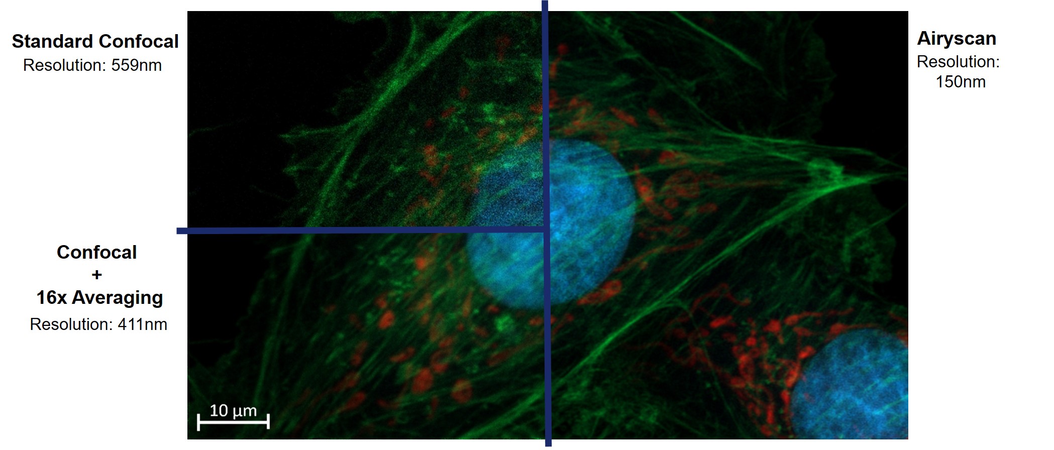
Comparison between Confocal and AiryScan imaging
Zen Connect
Zen connect is a feature present on all the Zeiss microscopes within the nmRC. This software makes it possible for data to be transferred directly between systems, keeping all information within a single project and directly correlating between systems where required. This can be done through may combinations but one of the most common applications is correlation of samples from CLSM to XB550.
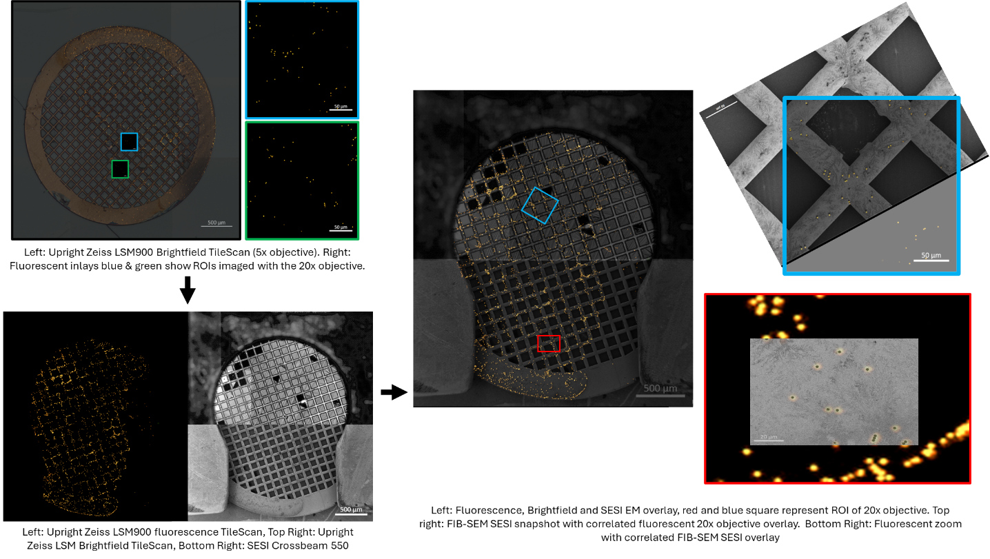
Zen connect example data showing correlation from Upright CLSM900 to the FIB-SEM XB550
Please contact nmRC CAT Suite team to discuss options and what would work for you.
- Zeiss Inverted CLSM 900 microscope with AiryScan 2 and maximum resolution of 120nm
- Zeiss Palm Micro Tweezers - optical tweezers for precise contact free cell manipulation
- Incubator stage for live-imaging
- Zen Connect
- 5x, 10x, 20x, 40x, 63x (oil), 100x (oil) objectives
Why use the CLSM900 Inverted:
- Best resolution (oil immersion) - ideal for smaller components e.g. organelles
- Requires thin glass coverslips/glass bottomed dishes
- Live imaging capabilities - visualise dynamic processes
- Micro tweezers for micromanipulation and rheology investigations
Images taken using each of the objectives on the inverted CLSM900, from 5x (air) to 100x (oil). Cells are Bovine foetal arterial endothelial cells stained with, DAPI (blue), Actin (green) and Mito Tracker (red).
- Zeiss Upright CLSM 900 microscope with AiryScan 2 and maximum resolution of 120 nm
- Linkam Cryo-Stage
- Room temperature stage
- Zen Connect
- 5x, 20x, 50x, 100x air objectives
Why use the CLSM900 Upright:
- Greater working distance - easier to image larger samples e.g. tissue
- Cryo-stage capabilities - image samples before and after vitrification
- Suitable for opaque containers or holders e.g. samples on TEM grids can be visualised with the upright
- Or containers not optimised for the inverted e.g. tissue culture plates and dishes
- Optimal perspective for correlation or comparison
Images taken using each of the objectives on the upright CLSM900, from 5x to 100x. Cells are Bovine foetal arterial endothelial cells stained with, DAPI (blue), Actin (green) and MitoTracker (red).
Zeiss LSM980 BFM Microscope
- Zeiss LSM 980 microscope AiryScan 2 with Multiplex Mode and maximum resolution of 120 nm
- Bruker NanoWizard V Scanning Head
- JPK CellHesion Head with 100 μm Z-range for large and strongly adhesive samples
- CytoSurge Fluid Force Microscope (FluidFM) which uses microsyringe, micropipette, and micro-trap FluidFM cantilevers
- Double-View optics which enables reflected and transmitted lights to be applied simultaneously
- PetriDish Heater - versatile holder for a wide range of 35 mm Petri dishes, including glass-bottom dishes
- Heats up to 60˚C
- Zen Connect
Why use the CLSM980 BFM:
- Perform high-precision AFM or Fluidic Force Microscopy (FluidFM) while simultaneously acquiring high-resolution, down to 120 nm, and time-resolved 3D structural imaging, up to 400 fps, using the AiryScan 2 with multiplex detection
- Obtain measurements including, morphology, mechanical properties, bioadhesion, nanorheology and protein-ligand interactions
- Micromanipulation using the FluidFM cantilever
- FluidFM allows for a localised delivery of (bio)chemicals to cell surfaces and tissues with video-rate monitoring of responses