Practical details and specimen requirements
Before commencing work it is worth noting a few basic requirements necessary in order to maximise the quality of the images produced on the ICS confocal.
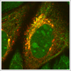 To take advantage of the high resolution oil/ water objective lenses cells must be grown/fixed on a No1.5 thickness glass coverslip. Lower resolution imaging of culture plates etc can be performed with 10x & 20x objectives.
Specimen thickness is an important factor. We can image to a depth of approximately 100um with the LSM510. Beyond this thickness results in loss of signal & resolution and so 2-Photon imaging is necessary.
 Fluorescent labels. We can image most conventional fluorescent dyes. Problems resulting from bleed through between channels can be removed by good experimental design, multi-tracking and emission fingerprinting with the META.
Images are saved to access style database. Data analysis and archiving can be performed by downloading the free Zeiss browser. Images can be converted to all well known formats if required. Work on the microscope can be in performed either by the CEO alone, with him or by yourself.
 Costs and details for future grant proposals can obtained from Tim Self. Use of the ICS imaging facilities are charged on an hourly, half day or daily basis.
Full training, costs and advice in all aspects of confocal microscopy are available from Tim Self : 0115 823 0090